
FIGURE
Fig 2
- ID
- ZDB-FIG-230526-56
- Publication
- Aljiboury et al., 2023 - Rab8, Rab11, and Rab35 coordinate lumen and cilia formation during zebrafish left-right organizer development
- Other Figures
- All Figure Page
- Back to All Figure Page
Fig 2
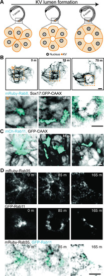
(A) Model depicting rosette, early lumen and late lumen stages of KV development being examined in live embryos represented in panels (B-D). (B-D) Representative images from live confocal videos of ectopically expressed mRuby-Rab8 (cyan, B), mCherry-Rab11 (cyan, C), endogenously tagged GFP-Rab11 (gray, cyan in merge, D) and ectopically expressed mRuby-Rab35 (gray, inverted gray in merge, D) localization in KV cells marked by GFP-CAAX (inverted gray, B) during KV lumen formation. Scale bar, 10μm. Refer to S2 Video. |
Expression Data
Expression Detail
Antibody Labeling
Phenotype Data
Phenotype Detail
Acknowledgments
This image is the copyrighted work of the attributed author or publisher, and
ZFIN has permission only to display this image to its users.
Additional permissions should be obtained from the applicable author or publisher of the image.
Full text @ PLoS Genet.