
- Title
-
A gap junction connexin is required in the vertebrate left-right organizer
- Authors
- Hatler, J.M., Essner, J.J., and Johnson, R.G.
- Source
- Full text @ Dev. Biol.
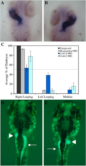
Cx43.4 is specifically required for L–R patterning. Dorsal views of 30 hpf embryos processed for in situ hybridization with a cardiac myosin light chain probe show normal looping of the heart tube towards the right in controls (A) and aberrant looping to the left in Cx43.4 morphants (B). (C) Quantification of heart looping in four independent knockdown experiments is represented as means ± SEM. The dotted line in all graphs shows the theoretical maximum value for randomization. (D) Ventral view of autofluorescence in the gut of a 5 dpf larva, specifically the gall bladder (arrowhead) and pancreas (arrow); compare with (E) the Cx43.4 morphant where these organs are reversed. |
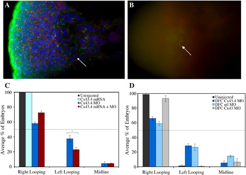
Cx43.4 is expressed in a spatiotemporal manner to be competent for KV function. Confocal fluorescence imaging (A) shows Cx43.4 labeling (red) in puncta that are distinct from the nuclei and cilia (green) of KV cells (arrow). Note also significant Cx43.4 labeling in the surrounding tail bud tissue. Grid-like confocal imaging (B) demonstrates Cx43.4 MO efficacy by the lack of staining (red), punctate or otherwise, in knockdown embryos. Cilia (green) of KV are labeled with tubulin antibodies and marked with an arrow as in (A). (C) Defects in heart laterality are partially rescued by coinjection of cx43.4 mRNA, ∗p = 0.04. (D) DFC-targeted injections of Cx43.4 MOs have a significant heart reversal phenotype. (C, D) Data are averages of three independent experiments and are represented as means ± SEM. EXPRESSION / LABELING:
PHENOTYPE:
|
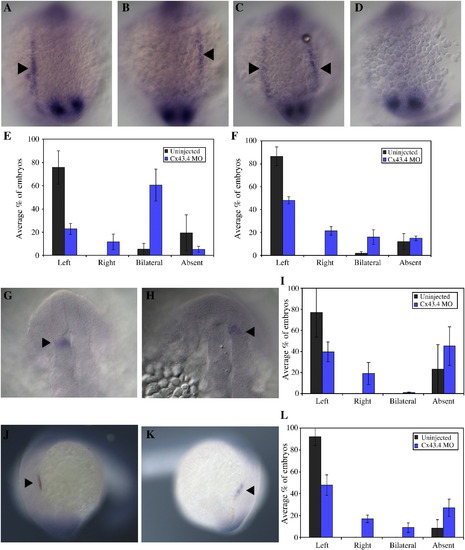
Molecular markers support a role for Cx43.4 in KV function. In situ hybridizations are used to probe for expression of genes in the L–R signaling cascade. (A–D) Twelve-somite Cx43.4 morphant embryos probed for spaw expression (arrowheads, throughout) show left, right, bilateral, and absent expression, respectively. spaw expression data are quantified (E) at 12 somites and (F) at 20 somites. Twenty-somite embryos probed with lefty1 show (G) normal expression in wild-type and (H) reversed expression in a DFC-targeted Cx43.4 knockdown embryo. (I) lefty1 data are summarized. (J) Twenty-somite embryos probed with lefty2 show a (K) reversal in the developing heart field of the Cx43.4 morphant. (L) Quantification of lefty2 expression was compiled from three independent experiments. Graphs in (E, F, I, L) represent means ± SEM from three independent experiments. EXPRESSION / LABELING:
PHENOTYPE:
|
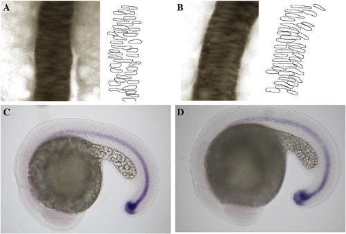
Midline integrity is maintained in Cx43.4 morphants. In situ hybridization with a ntl probe (A–D). (A) Ten-somite stage uninjected and (B) Cx43.4 morphant embryos both have an intact, continuous midline. For clarity, individual nuclei were outlined in Photoshop (right of A and B). (C) Twenty-somite stage control and (D) Cx43.4-deficient embryos express ntl along the midline. EXPRESSION / LABELING:
PHENOTYPE:
|
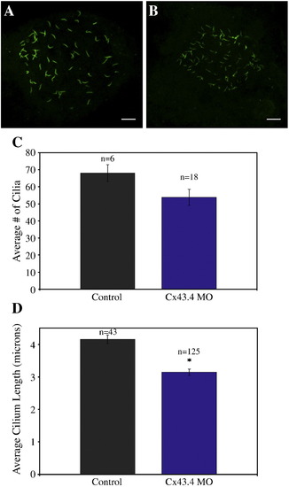
KV cells are specified in Cx43.4 morphants, though ciliary defects exists. Confocal fluorescence imaging (A, B) of KV cilia labeled with acetylated tubulin antibodies. Scale bar = 10 microns. (A) Uninjected, control embryos have well-developed cilia in a circular pattern, while (B) Cx43.4 morphants have disorganized, smaller ciliated KVs. Multiple fluorescence images of 10-somite stage KVs labeled with acetylated tubulin antibiodies were analyzed using ImageJ software (C, D) and data were plotted as means ± SEM. (C) The average number of cilia per KV was similar between uninjected and Cx43.4 morphant embryos, p = 0.13. (D) The average cilium in Cx43.4 morphant embryos was significantly shorter than a wild-type cilium, ∗p = 3 x 10- 7. EXPRESSION / LABELING:
PHENOTYPE:
|
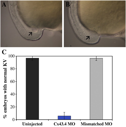
KV lumen formation is impaired in Cx43.4 morphants. DIC microscopy (A, B) of 10-somite stage embryos. (A) KV inflation is observed in control embryos, while (B) Cx43.4 morphants do not have detectable KVs (arrows in A and B point to KV location). (C) The percentage of embryos with normal KVs as analyzed by DIC microscopy from three independent experiments are represented as means ± SEM. PHENOTYPE:
|
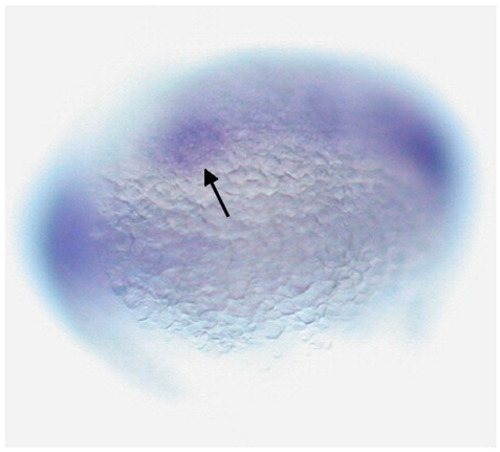
Cx44.2 is expressed in KV. Ventral view of a 10-somite stage embryo processed by for in situ hybridization with cx44.2 antisense probes shows cx44.2 expression in KV (arrow). EXPRESSION / LABELING:
|
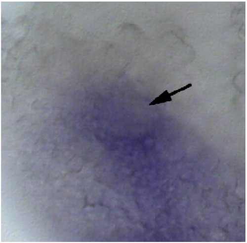
Cx43.4 is expressed in KV. In situ hybridization analysis shows that cx43.4 transcripts are expressed in KV (arrow) of a 10-somite stage embryo. EXPRESSION / LABELING:
|

Cx43.4 fluorescein-tagged MOs are targeted to the DFCs. (A) Dorsal view of a representative shield stage embryo showing fluorescently labeled Cx43.4 MOs in clusters of DFCs (arrowheads). (B) Cx43.4 MOs injected at the mid-blastula stage were restricted to the yolk at 24 hpf. |

Unillustrated author statements EXPRESSION / LABELING:
PHENOTYPE:
|
Reprinted from Developmental Biology, 336(2), Hatler, J.M., Essner, J.J., and Johnson, R.G., A gap junction connexin is required in the vertebrate left-right organizer, 183-191, Copyright (2009) with permission from Elsevier. Full text @ Dev. Biol.