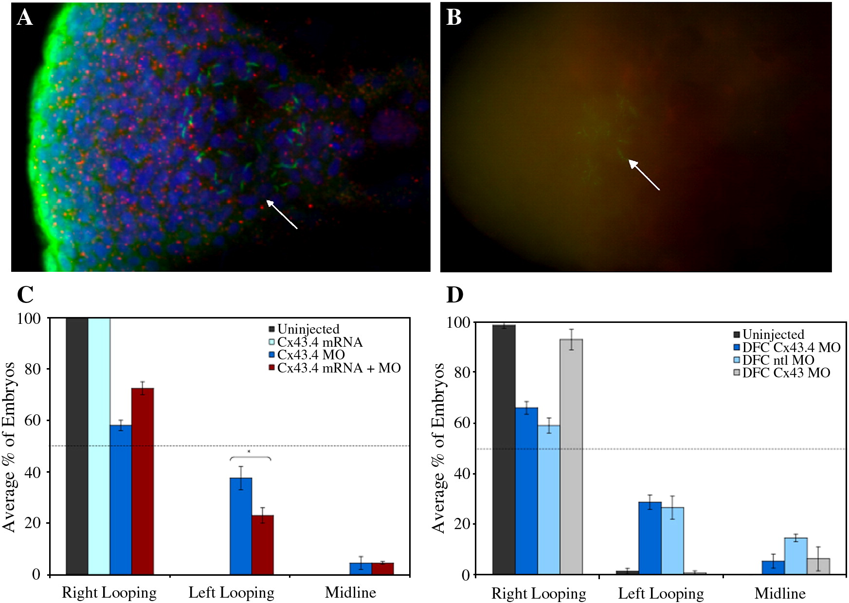
Fig. 2 Cx43.4 is expressed in a spatiotemporal manner to be competent for KV function. Confocal fluorescence imaging (A) shows Cx43.4 labeling (red) in puncta that are distinct from the nuclei and cilia (green) of KV cells (arrow). Note also significant Cx43.4 labeling in the surrounding tail bud tissue. Grid-like confocal imaging (B) demonstrates Cx43.4 MO efficacy by the lack of staining (red), punctate or otherwise, in knockdown embryos. Cilia (green) of KV are labeled with tubulin antibodies and marked with an arrow as in (A). (C) Defects in heart laterality are partially rescued by coinjection of cx43.4 mRNA, ∗p = 0.04. (D) DFC-targeted injections of Cx43.4 MOs have a significant heart reversal phenotype. (C, D) Data are averages of three independent experiments and are represented as means ± SEM.
Reprinted from Developmental Biology, 336(2), Hatler, J.M., Essner, J.J., and Johnson, R.G., A gap junction connexin is required in the vertebrate left-right organizer, 183-191, Copyright (2009) with permission from Elsevier. Full text @ Dev. Biol.

