
Fig. 4
|
Fig. 4
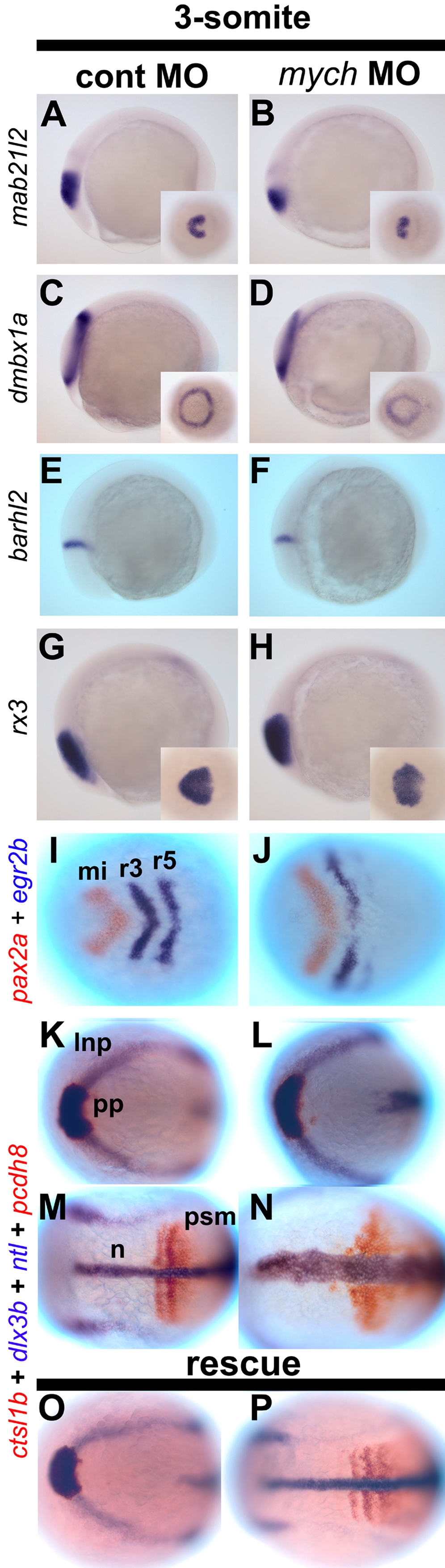
Mych MO affects multiple regions at the 3-somite stage.
A–P. Lateral views (A–H) and dorsal views (I–P) of control MO-injected (A,C,E,G,I,K,M) and mych MO-injected embryos (B,D,F,H,J,L,N). Mab21l2 (A–B′) as eye anlage and midbrain marker; dmbx1a (C–D′) as eye territory and tectum marker; barhl2 (E,F) as diencephalon marker; rx3 as eye field marker. (I–J) double label with pax2a (red) and egr2b (blue). (K–P). Embryos were examined using four probes, ctsl1b (red), dlx3b (blue), ntl (blue), and pcdh8 (red). (O–P) mych MO-induced defects were rescued by co-injection with mych mRNA. lnp, lateral neural plate; mi, midbrain; n, notochord; pp, prechordal plate; r, rhombomere; psm, pre somitic mesoderm.