
Fig. 4
|
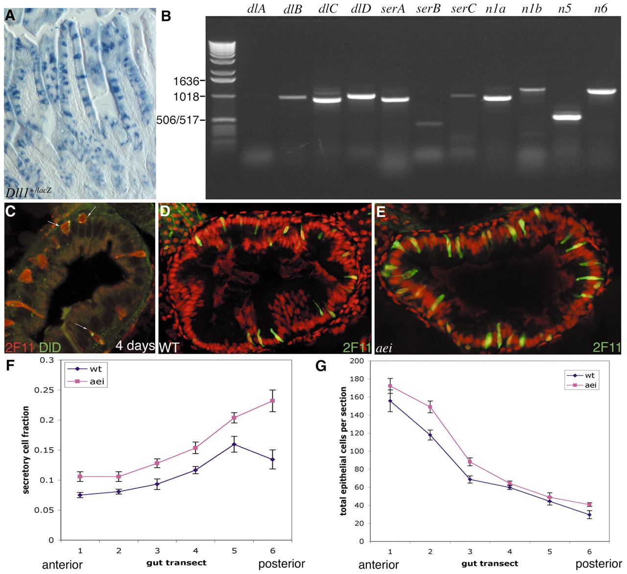
Fig. 4 Secretory cells express the Notch ligand Delta and their numbers are increased in aeiAR33, a mutant lacking DeltaD. (A) Intestine of adult mouse heterozygous for a lacZ knock-in at the Delta1 (Dll1) locus. ß-Galactosidase activity (blue) is a reporter for present or past expression of Delta1 expression; the stain is seen in scattered cells, many of which can be clearly identified as goblet cells. (B) Expression of Notch pathway components in zebrafish gut, analysed by RT-PCR. deltaC and deltaD are strongly expressed in the gut, while deltaA and deltaB are not. (C) DeltaD (zdd-2 immunostain in green, arrows) is visible in a subset of secretory cells (2F11, red) in the wild-type zebrafish intestine at 4 days. (D,E) Sections of intestinal bulb of 5-day-old wild-type larvae (D) and aeiAR33 homozygotes (E) stained with 2F11; TOPRO-3 nuclear stain is red. (F) The proportion of 2F11-positive cells is increased all along the length of the gut in aeiAR33 when compared with wild type; data points show mean and s.e.m. from counts of sections of 12 larvae of each genotype. (G) Total numbers of epithelial cells per section for the same set of specimens.