|
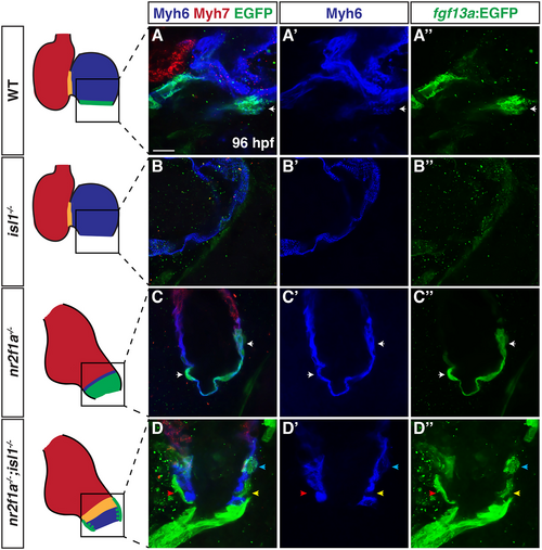
Cells in the venous atria of nr2f1a;isl1 mutants lack Myh6. (A-D″) Optical sections of the venous pole of wild-type, isl1−/−, nr2f1a−/− and nr2f1a−/−;isl1−/− hearts at 96 hpf stained for Myh6 (blue), Myh7 (red) and fgf13a:EGFP (green). In the schematics, Myh7 (red), Myh6 (blue), Myh6 + Myh7 (orange) and fgf13a:EGFP (green) are shown. Overlap of Myh6 and fgf13a:EGFP in myocardium in A-A″ and C-C″ (white arrows). (D-D″) A fgf13a:EGFP+ cell in a gap of Myh6 (yellow arrows), a fgf13a:EGFP+ cell on the outside of the atrial myocardial wall (red arrows) and a fgf13a:EGFP+/Myh6+ cell in the atrial myocardial wall (blue arrows). Scale bar: 10 μm. Wild type (n=9), nr2f1a−/− (n=11), isl1−/− (n=4) and nr2f1a−/−;isl1−/− (n=13).
|