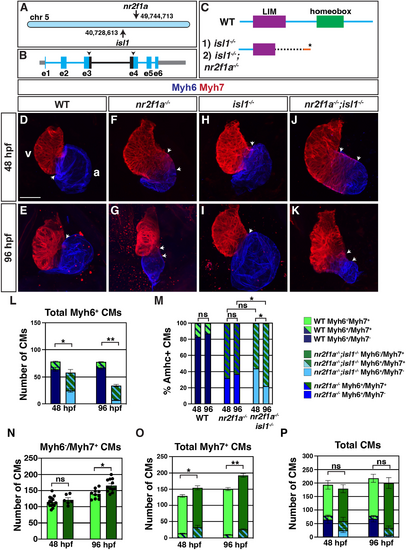
nr2f1a;isl1 mutants have reduced atrial cardiomyocytes. (A) Schematic of nr2f1a and isl1 loci on chromosome 5. (B) Schematic of deletion in isl1 locus. Location of guide RNAs and the boundaries of deletion (black arrowheads). (C) Schematic of predicted consequences of genomic deletion on Isl1 protein in isl1 mutants and nr2f1a;isl1 mutants. (D-K) Immunohistochemistry for Myh6 (blue) and Myh7 (red) in the wild-type, nr2f1a−/−, isl1−/− and nr2f1a;isl1−/− hearts at 48 and 96 hpf. Overlap of Myh6 and Myh7 in the atrioventricular canal is indicated (white arrows). v, ventricle; a, atrium. Scale bar: 25 μm. 48 hpf: wild type (n=11), nr2f1a−/− (n=4), isl1−/− (n=13), nr2f1a−/−;isl1−/− (n=7); 96 hpf: wild type (n=16), nr2f1a−/− (n=11), isl1−/− (n=8), nr2f1a−/−;isl1−/− (n=8). Individual channels with the merged images shown in D-K are presented in Fig. S1A-H. (L) Quantification of total Myh6+ (Myh6+/Myh7−+Myh6+/Myh7+) cardiomyocytes (CMs). (M) Proportions of Myh6+/Myh7− and Myh6+/Myh7+ CMs at 48 and 96 hpf. Proportion of CMs for nr2f1a mutants are derived from Martin et al. (2023). (N-P) Quantification of Myh6−/Myh7+ (Myh7-only), total Myh7+ (Myh6+/Myh7+ and Myh6−/Myh7+) and total CMs. 48 hpf: wild type (n=15) and nr2f1a−/−;isl1−/− (n=6); 96 hpf: wild type (n=10) and nr2f1a−/−;isl1−/− (n=12). *P=0.05-0.001; **P<0.001 (ANOVA with multiple comparisons in L,N-P; Fisher's exact test in M).
|
