|
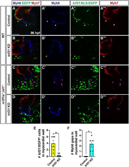
tcf21 is required to promote epicardial cell gene expression in nr2f1a;isl1 mutant atria. (A-D‴) Optical sections of immunohistochemistry for Myh6 (blue), Myh7 (red) and tcf21:NLS-EGFP (green) in control and tcf21 knockdown (KD) wild-type and nr2f1a−/−;isl1−/− hearts. (A-A‴) Epicardial cells (ECs) on the ventricle (white arrowheads). (B-B‴) The lack of ECs on the ventricle (yellow arrowheads). (C-C‴) tcf21:NLS-EGFP+/Myh6− cells in myocardial wall (magenta arrowheads). (D-D‴) Gaps in Myh6 within the myocardial wall (blue arrowheads). Scale bar: 25 μm. Control: wild type (n=9) and nr2f1a−/−;isl1−/− (n=6); tcf21 KD: wild type (n=9) and nr2f1a−/−;isl1−/− (n=5). (E) Quantification tcf21:NLS-EGFP+ cells within the control and tcf21 KD nr2f1a−/−;isl1−/− hearts. *P=0.0106 (two-tailed Welch's t-test). (F) Quantification of gaps in Myh6+ myocardial wall of control and tcf21 KD nr2f1a−/−;isl1−/− hearts. *P=0.0161 (two-tailed Welch's t-test).
|