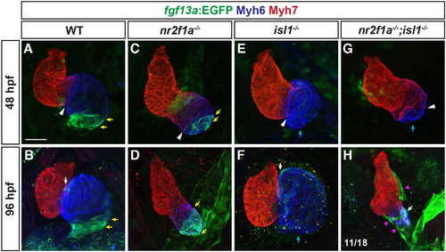
nr2f1a;isl1 mutants lack pacemaker cardiomyocytes. (A-H) Immunohistochemistry for Myh6 (blue), Myh7 (red) and fgf13a:EGFP (green) in hearts of wild-type, nr2f1a mutant, isl1 mutant and nr2f1a;isl1 mutant embryos at 48 and 96 hpf. The extent of Myh7 expression within the hearts (white arrowheads), the limits of fgf13a:EGFP expression within the heart (yellow arrows), the lack of fgf13a:EGFP expression (blue arrows), the AV node (white arrow) and Fgf13a:EGFP+ cells on the surface of the heart (magenta arrows) are shown. Representative image in H depicts the phenotype observed in the majority of nr2f1a;isl1 mutant hearts (11/18) for this experiment. The 96 hpf wild-type heart in B is also presented in Fig. S3A for comparison to the phenotype observed in the minority of nr2f1a;isl1 mutant hearts (7/18) presented in Fig. S3B. Scale bar: 25 μm. 48 hpf: wild type (n=15), nr2f1a−/− (n=9), isl1−/− (n=13) and nr2f1a−/−;isl1−/− (n=12); 96 hpf: wild type (n=13), nr2f1a−/− (n=11), isl1−/− (n=8) and nr2f1a−/−;isl1−/− (n=18).
|
