|
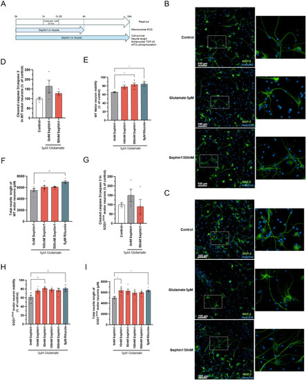
Sephin1 protects primary WT and SOD1G93A rat motor neurons against glutamate injury. (A) Schematic of the experiment on primary WT and SOD1G93A rat motor neurons. (B) Representative pictures of primary WT rat motor neurons treated with glutamate 5 μM for 20 min in presence of DMSO (vehicle) or Sephin1 500 nM 24 h after glutamate intoxication. Motor neurons are labeled with the neuronal marker, MAP-2. Right panel: enlargement of motor neurons (C) Representative pictures of primary SOD1G93A rat motor neurons treated with glutamate 5 μM for 20 min in presence of DMSO (vehicle) or Sephin1 50 nM 24 h after glutamate intoxication. Motor neurons are labeled with the neuronal marker, MAP-2. Right panel: enlargement of motor neurons (D) Cleaved caspase 3 level measured 24 h after glutamate intoxication in primary WT rat motor neurons culture treated with glutamate 5 μM for 20 min in presence of DMSO (vehicle) or Sephin1 50 nM. (E) Cell viability in glutamate intoxicated primary WT rat motor neurons treated for 24 h with DMSO (Vehicle), Sephin1, or riluzole. (F) Total neurite length of primary WT rat motor neurons treated for 24 h with DMSO (Vehicle), Sephin1 or riluzole 24 h after glutamate intoxication. (G) Cleaved caspase 3 level measured 24 h after glutamate intoxication in primary SOD1G93A rat motor neurons culture treated with glutamate 5 μM for 20 min in presence of DMSO (vehicle) or Sephin1 50 nM 24 h. (H) Cell viability in glutamate intoxicated primary SOD1G93A rat motor neurons treated for 24 h with DMSO (Vehicle), Sephin1 or riluzole. (I) Total neurite length of primary WT rat motor neurons treated for 24 h with DMSO (Vehicle), Sephin1 or riluzole 24 h after glutamate intoxication. *P < 0.05, **P < 0.01 versus 0 nM Sephin1 Kruskal-Wallis test followed by Dunn’s multiple comparison test; mean ± SEM n = 4–6 replicates per condition. Source data are available for this figure.
|