
Fig. Ext 4
- ID
- ZDB-FIG-250428-9
- Publication
- Thiruppathy et al., 2025 - Repurposing of a gill gene regulatory program for outer ear evolution
- Other Figures
- All Figure Page
- Back to All Figure Page
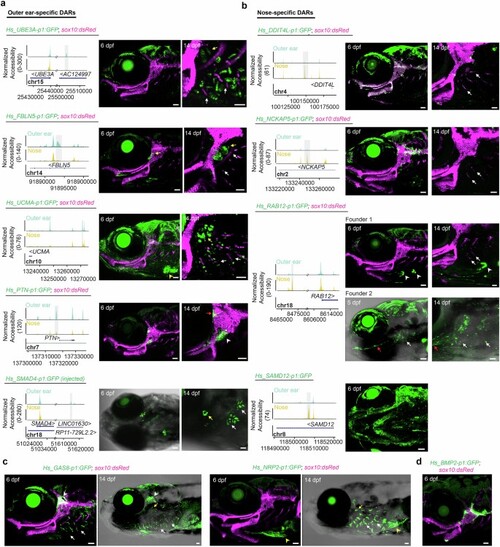
a,b, Genome coverage plots and zebrafish transgenic activity of human outer-ear-specific and nose-specific cartilage DARs. Except for Hs_SMAD4-p1:GFP-injected fish, all transgenic images are of stable lines. Lateral views of the head are shown at 5-6 dpf, and higher magnification views of the hyomandibular (hyaline) cartilage and gills are shown at 14 dpf. sox10:dsRed labels all cartilage. White arrows indicate the gill filaments and yellow arrows the gill-like pseudobranch. For Hs_UCMA-p1:GFP, a yellow arrowhead indicates heart expression at 6 dpf. For Hs_PTN-p1:GFP, a white arrowhead indicates mesenchymal expression near the hyomandibula and a red arrow labels a subset of hyomandibular-otic joint cells. For Hs_RAB12-p1:GFP, Founder 1 shows expression at the tips of the branchiostegal ray bones (arrowheads) and Founder 2 has weak and variable expression in the gill region (white arrows) in addition to the interopercular-mandibular ligament (red arrow). c, Additional images related to Fig. 3b,d show specificity of Hs_GAS8-p1:GFP for the gill filaments at 6 and 14 dpf and Hs_NRP2-p1:GFP for the gill filaments at 14 dpf. White arrowheads indicate labeling of trigeminal ganglia by Hs_GAS8-p1:GFP, and yellow arrowheads seventh arch mesenchyme labeling by Hs_NRP2-p1:GFP. d, Hs_BMP2-p1:GFP labels subsets of otic, hyomandibular, and ceratohyal hyaline cartilage at 6 dpf. Scale bars, 50 µm. |