
Fig. Ext. 9
- ID
- ZDB-FIG-250428-14
- Publication
- Thiruppathy et al., 2025 - Repurposing of a gill gene regulatory program for outer ear evolution
- Other Figures
- All Figure Page
- Back to All Figure Page
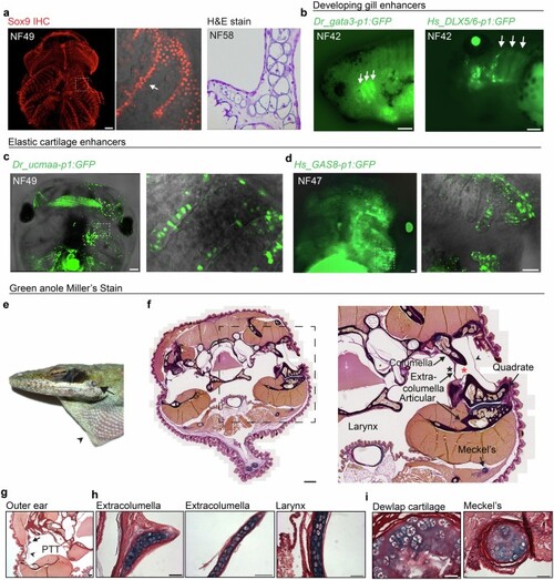
a, Whole-mount Sox9 immunostaining of cartilage in an NF49 Xenopus laevis tadpole head (ventral view, anterior up). Magnified boxed region highlights gill filter cartilage (arrow). H&E stain on a comparable section through the gill filters of an NF58 tadpole shows cellular cartilage (reproduced from Figure S2e in ref. 23). b, White arrows highlight gata3-p1:GFP (left) and Hs_DLX5/6-p1:GFP (right) activity in the developing gills of NF42 tadpoles, which persisted up to late tadpole stages NF47-49. c,d, Elastic cartilage enhancers Dr_ucmaa-p1:GFP (c) and Hs_GAS8-p1:GFP (d) drove reporter activity in gill filter cartilage (magnified boxed regions) in NF47-49 tadpoles. No expression was seen in the early developing gills at NF42 from either construct. Expression is unilateral due to injection of one cell at the two-cell stage, with non-specific muscle fluorescence. e, Adult head of a green anole (Anolis carolensis) showing the shallow outer ear (arrow) and dewlap (arrowhead). f-i, Miller’s Stain of frontal sections through the green anole head. Boxed magnified region in f shows anatomy surrounding the pharyngotympanic tube (black asterisk). The extracolumella, attached to the columella, extends processes into the pharyngotympanic tube and towards the tympanic membrane (arrowhead). The short outer ear canal (red asterisk) sits directly above the jaw joint, comprising the articular and quadrate bones, and Meckel’s cartilage is ventral to the jaw joint. Compared to the extracolumella permanent cartilage, the columella has completely ossified. The pharyngotympanic tube (PTT) and tympanic membrane (arrowhead) supported by the extracolumella (arrow) are highlighted in g. Higher magnification images contrast the cellular nature and strong Miller’s stain of the extracolumella and laryngeal elastic cartilages (h) with the permanent hyaline dewlap and Meckel’s cartilages (i). Scale bars, 200 µm (a, c,f), 50 µm (b,d,g-i). |