
Fig. Ext 5
- ID
- ZDB-FIG-250428-10
- Publication
- Thiruppathy et al., 2025 - Repurposing of a gill gene regulatory program for outer ear evolution
- Other Figures
- All Figure Page
- Back to All Figure Page
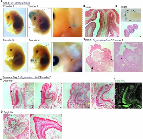
a, Representative images from whole-mount X-gal staining of Dr_ucmaa-p1:lacZ mice at E15.5 from 4 independent founder lines. Black arrows highlight outer-ear staining observed in 3 of 4 founders. We also observed digit expression in all 4 founders, nasal expression in Founders 1 and 2, and variable forebrain and weak hair follicle expression in Founder 2. b, Horizontal section through nose of Founder 2 shows activity in the olfactory epithelium (white arrowheads indicate rare cells) but not nasal hyaline cartilage (C). c, Magnified boxed region from Founder 4 shows positive staining in the mouse digit tips. Transverse section reveals expression in collateral ligaments surrounding the digit cartilage (C). d, Frontal section through Founder 1 shows expression in the outer ears (black arrows) but absence of expression in epiglottis (green arrow) and surrounding laryngeal hyaline cartilages (yellow arrowhead). e,g, X-gal staining of horizontal cryosections from the outer ear (e) and epiglottis (g) of Founder 2 P0 pups, showing activity in the ear canal cartilages (arrows) in boxed region of the third panel, but not in elastic cartilage of the pinna (black arrowheads) or epiglottis (boxed region in g). Hyaline cartilages of the middle ear and larynx (yellow arrowheads) are also negative. f, Sox9 immunostaining on adjacent sections confirms that the positive tissue of the outer ear is cartilage. All X-gal stained sections are counterstained with nuclear fast red. Scale bars, 50 µm (a-d), 100 µm (e-g). |