|
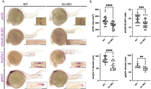
Further analysis of pronephros segment development in fxn-deficient zebrafish embryos confirms reductions in several cell populations. (A) WISH using the probes wt1b, trim35-30-201, kcnj1a.1, and gata3 which mark the podocytes, multiciliated cells (MCCs), distal early and distal late domains respectively. Fxn morphants displayed a significant reduction in podocytes, reduction in MCC number, and reduced distal domains results with this set of markers, independently recapitulating the results shown in Figure 3. Scale bars = 50 um. Each boxed lateral region in the embryos corresponds to the inset, which shows a dorsal view of that corresponding area, with the exception of the gata3 panels which show lateral views in the corresponding inset. (B) Unpaired t-tests demonstrating significantly affected pronephric domains in the fxn morphants. **p < 0.005, ***p < 0.0005, ****p < 0.0001.
|