IMAGE

FIGURE 4

ID
ZDB-IMAGE-250109-297
Genes
Source
Figures for Ercanbrack et al., 2024
Image
Figure Caption

FIGURE 4

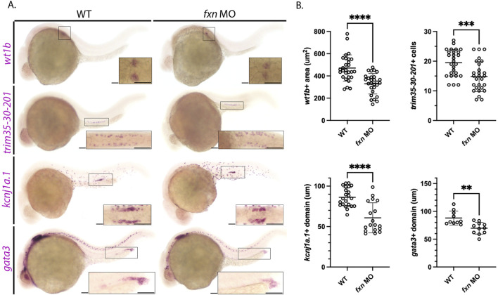
Further analysis of pronephros segment development in fxn-deficient zebrafish embryos confirms reductions in several cell populations. (A) WISH using the probes wt1b, trim35-30-201, kcnj1a.1, and gata3 which mark the podocytes, multiciliated cells (MCCs), distal early and distal late domains respectively. Fxn morphants displayed a significant reduction in podocytes, reduction in MCC number, and reduced distal domains results with this set of markers, independently recapitulating the results shown in Figure 3. Scale bars = 50 um. Each boxed lateral region in the embryos corresponds to the inset, which shows a dorsal view of that corresponding area, with the exception of the gata3 panels which show lateral views in the corresponding inset. (B) Unpaired t-tests demonstrating significantly affected pronephric domains in the fxn morphants. **p < 0.005, ***p < 0.0005, ****p < 0.0001.

Figure Data
Acknowledgments
This image is the copyrighted work of the attributed author or publisher, and ZFIN has permission only to display this image to its users. Additional permissions should be obtained from the applicable author or publisher of the image. Full text @ Front Cell Dev Biol