FIGURE

Figure S6.

ID
ZDB-FIG-241021-33
Publication
El-Daher et al., 2024 - Microglia are essential for tissue contraction in wound closure after brain injury in zebrafish larvae
Other Figures
All Figure Page
Back to All Figure Page
Figure S6.

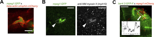
Microglia possess the molecular machinery to exert forces, and accumulation leads to increased GFAP detectability.

(A) Confocal micrograph of a microglial cell in the optic tectum, which expresses both mpeg1:GFP and beta-actin:utrophin-mCherry, illustrating that the F-actin filaments accumulate along the exterior membrane of the microglia after stab injury. (B) Microglial cell (mpeg1:GFP) immunofluorescently labelled for non-muscular myosin II, the target for blebbistatin. The cell shows notable accumulation of myosin II. (C) Horizontal section of the tectum at 12 h post-injury is shown, together with a density plot for GFP fluorescence. Astrocytic processes labelled by her4.3:GFP-F show increased fluorescence next to the accumulation of microglia in the tectal neuropil. The scale bar in (A, B) represents 10 μm.

Expression Data

Expression Detail
Antibody Labeling
Phenotype Data

Phenotype Detail
Acknowledgments
This image is the copyrighted work of the attributed author or publisher, and ZFIN has permission only to display this image to its users. Additional permissions should be obtained from the applicable author or publisher of the image. Full text @ Life Sci Alliance