
FIGURE
Figure S7.
- ID
- ZDB-FIG-241021-34
- Publication
- El-Daher et al., 2024 - Microglia are essential for tissue contraction in wound closure after brain injury in zebrafish larvae
- Other Figures
- All Figure Page
- Back to All Figure Page
Figure S7.
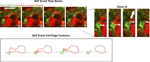
Utrophin labelling is enriched at sites of astrocyte pulling. (Upper left) Fast time-lapse imaging sequence on a Tg(her4.3:GFP-F); Tg(beta-actin:utrophin-mCherry) larva showing an SAT process. The white arrow points out the adhesion event. (Right) Cropped sequence from upper left. White arrows point to the astrocytic node pulled by microglial protrusion. The dashed line indicates the initial position. (Lower left) Outline of microglia (red) and astrocytic structures (green) from images in the panel above. Arrows show the direction of displacement. Scale bar: 10 μm. |
Expression Data
Expression Detail
Antibody Labeling
Phenotype Data
Phenotype Detail
Acknowledgments
This image is the copyrighted work of the attributed author or publisher, and
ZFIN has permission only to display this image to its users.
Additional permissions should be obtained from the applicable author or publisher of the image.
Full text @ Life Sci Alliance