FIGURE

Figure 7.

ID
ZDB-FIG-241021-35
Publication
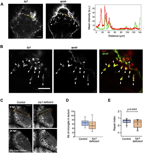
El-Daher et al., 2024 - Microglia are essential for tissue contraction in wound closure after brain injury in zebrafish larvae
Other Figures
All Figure Page
Back to All Figure Page
Figure 7.

Microglial action relies on lcp1.

(A) Expression of lcp1 and apoeb as detected by hybridisation chain reaction fluorescence in situ hybridisation in injured larvae at 6 hpi. The intensity profiles shown on the graph (red: lcp1; green: apoeb) were measured along the orange lines shown in the images. (B) Zoomed-in hybridisation chain reaction fluorescence in situ hybridisation images for lcp1a (green) and apoeb (red), showing an almost complete overlap of patterns. Arrows point to lcp1+/apoeb+ cells at the lesion site. (C) Representative images of wound closure in control and haCR-injected animals are shown; orange dashed lines: injury site; white dashed lines: wound delimitation in the PVZ. (D) Boxplot representation of the repair index in all haCR-injected animals (P from an unpaired t test). (E) Boxplot representation of the number of microglia in the optic tectum at 24 hpi in haCR-injected animals. All t tests were non-significant (P > 0.05). For all experiments, n = 4 for control-injected, and n = 10 for haCR-injected animals. All scale bars represent 50 μm.

Expression Data

Expression Detail
Antibody Labeling
Phenotype Data

Phenotype Detail
Acknowledgments
This image is the copyrighted work of the attributed author or publisher, and ZFIN has permission only to display this image to its users. Additional permissions should be obtained from the applicable author or publisher of the image. Full text @ Life Sci Alliance