
Fig. 5
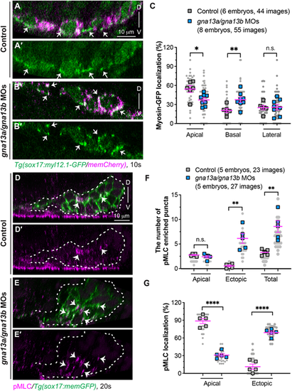
Gα13 controls spatial actomyosin activity to regulate endoderm convergence. (A-C) Myosin dynamics in the indicated embryos at 10 ss. (A-B′) Confocal images of xz planes showing the localization of Myl12.1-GFP in mem-mCherry-labelled endodermal cells (magenta). Arrows indicate enriched Myl12.1-GFP labeled puncta. (C) The frequencies of Myl12.1-GFP labeling in the apical, basal and lateral region of endodermal cells in xz planes. (D-G) Expression of pMLC, detected by whole-mount immunostaining, in the indicated embryos at 20 ss. (D-E′) Confocal images of xz planes showing the localization of pMLC (magenta) in memGFP-labelled endodermal cells. Arrows indicate pMLC-labeled puncta. White dashed lines outline the endoderm. (F,G) Distribution of pMLC expression. (F) The average number of pMLC-labeled puncta in the apical and ectopic regions, as well as the total number. (G) The frequencies of pMLC-labelled puncta in the apical and ectopic regions in the indicated embryos. Data from all embryos (squares) and all XZ images (gray circles) are superimposed, with the number of xz images and embryos indicated. Data are mean±s.e.m. n.s. (not significant), P>0.05; *P<0.05; **P<0.01; ****P<0.0001 (unpaired, two-tailed Student's t-test). D, dorsal; V, ventral. |