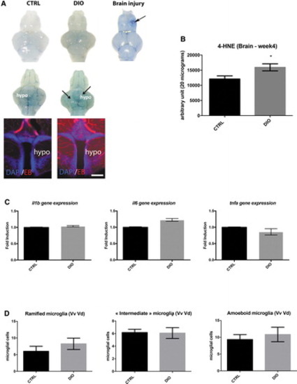
DIO impact on BBB leakage, cerebral oxidative stress, and neuroinflammation. (A) First row: dorsal view pictures of the brain from DIO model CTRL and DIO fish. Second row: ventral view pictures of the brain from DIO model CTRL and DIO fish (n = 3–6 brains). Note the barely blue stained brain parenchyma of DIO-treated fish. Third column: positive control showing Evans blue extravasation in the injured telencephalon (arrows). In (A), hypothalamic vibratome section showing Evans blue staining (Red) extravasation in the parenchyma. (B) Graph showing dot-blot quantification of 4-HNE staining in the brains of CTRL and DIO fish (n = 4). (C) Proinflammatory cytokines (il1β, il6, and tnfα) cerebral gene expression in CTRL and DIO conditions (n = 3 pools of 2 brains). (D) Counting of ramified, “intermediate,” and ameboid microglia in the ventral part of the telencephalon (Vv Vd region) in both CTRL and DIO fish (n = 6). Bar graph: SEM. Student's t-test: *p < 0.05. Scale bar: 1 mm for whole brain picture and 75 μm for hypothalamic sections. BBB, blood–brain barrier; n, number of fish; Vd, dorsal nucleus of ventral telencephalic area; Vv, ventral nucleus of ventral telencephalic area. Color images are available online.
|
