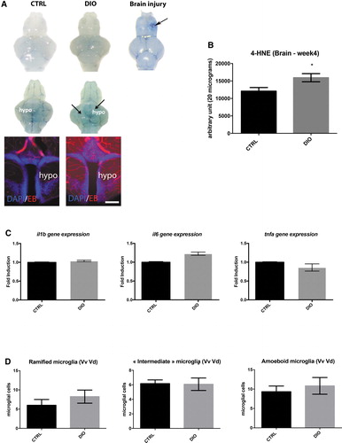
Fig. 3 DIO impact on BBB leakage, cerebral oxidative stress, and neuroinflammation. (A) First row: dorsal view pictures of the brain from DIO model CTRL and DIO fish. Second row: ventral view pictures of the brain from DIO model CTRL and DIO fish (n = 3–6 brains). Note the barely blue stained brain parenchyma of DIO-treated fish. Third column: positive control showing Evans blue extravasation in the injured telencephalon (arrows). In (A), hypothalamic vibratome section showing Evans blue staining (Red) extravasation in the parenchyma. (B) Graph showing dot-blot quantification of 4-HNE staining in the brains of CTRL and DIO fish (n = 4). (C) Proinflammatory cytokines (il1β, il6, and tnfα) cerebral gene expression in CTRL and DIO conditions (n = 3 pools of 2 brains). (D) Counting of ramified, “intermediate,” and ameboid microglia in the ventral part of the telencephalon (Vv Vd region) in both CTRL and DIO fish (n = 6). Bar graph: SEM. Student's t-test: *p < 0.05. Scale bar: 1 mm for whole brain picture and 75 μm for hypothalamic sections. BBB, blood–brain barrier; n, number of fish; Vd, dorsal nucleus of ventral telencephalic area; Vv, ventral nucleus of ventral telencephalic area. Color images are available online.
Image
Figure Caption
Figure Data
Acknowledgments
This image is the copyrighted work of the attributed author or publisher, and
ZFIN has permission only to display this image to its users.
Additional permissions should be obtained from the applicable author or publisher of the image.
Full text @ Zebrafish

