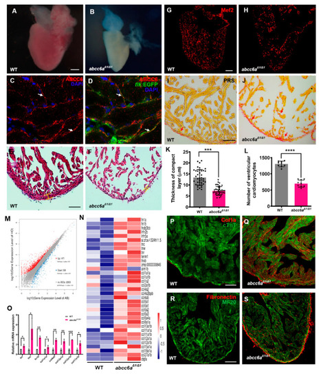
abcc6aΔ1/Δ1 mutation upregulates extracellular matrix genes and leads to fibrotic adult hearts. (A,B) Whole-mounted photographs of adult heart at 8 mpf showing cardiac shrinkage and fibrosis phenotypes in abcc6aΔ1/Δ1 (B), compared with WT sibling heart (A). (C,D) Abcc6 protein (red) recognized with anti-Abcc6 antibody overlaps with endocardial cells marked by flk:EGFP (green) in adult Tg(flk:EGFP) heart. Arrows in (C,D) point to endocardial cells expressing Abcc6. (E,F) Hematoxylin-eosin (HE) staining exhibits thinner compact layer and fewer myocardial cells in abcc6aΔ1/Δ1 heart (F), compared to WT sibling hearts (E). (G,H) Representative Picrosirius Red staining of heart sections (yellow for cardiomyocyte fiber, red for collagen) shows superfluous collagen and elastin deposition residing in a compact layer and trabeculae in abcc6aΔ1/Δ1 adult hearts (H), compared to WT hearts (G). (I,J) Immunofluorescent section images of abcc6aΔ1/Δ1 mutant hearts (J) and WT siblings (I), stained with anti-Mef2 antibody. (K) Quantitative analysis shows a thinner compact layer thickness in abcc6aΔ1/Δ1 adult heart (n = 10, 4 measuring points per sample), compared with WT sibling heart (n = 10, 4 measuring points per sample). *** p < 0.001, Student’s t-test (unpaired, two-tailed). (L) Quantification of the number of cardiomyocyte cells in the same area of WT (n = 9) and abcc6aΔ1/Δ1 mutants (n = 9). **** p < 0.0001, Student’s t-test (unpaired, two-tailed). (M) Volcano plots showing differentially expressed genes. 1471 genes are increased in abcc6aΔ1/Δ1 mutant hearts and 599 genes expression are reduced, compared to WT hearts. (N) Heat map indicates genes upregulated in abcc6aΔ1/Δ1 mutant hearts, compared to WT hearts (higher expression in red, lower expression in blue). FC > 1.5, p < 0.05. (O) qPCR analyses of tenascin family genes (tnr and tnc) and fibronectin b and collagen family genes (col4a5, col12a1a, col12a1b and col17a1b) in hearts extracted from abcc6aΔ1/Δ1 and abcc6+/+ animals. Data presented as mean ± SEM, n = 3, * p < 0.05, ** p < 0.01, *** p < 0.001, Student’s t-test (unpaired, two-tailed). (P,Q) Immunostaining analyses reveal a robust collagen I in abcc6a mutant hearts (Q), compared to that in WT hearts (P), stained with anti-Col1a (red) and anti-cTnT antibodies (green). (R,S) Immunostaining analyses show pervading Fibronectin in compact and trabecular layers in abcc6aΔ1/Δ1 hearts (S) compared to WT hearts (R), stained with anti-Fibronectin (red) and anti-MF20 antibodies (green). Scale bar: 100 μm (A,B; G,H; R,S); 50 μm (E,F); 20 μm (P,Q).
|
