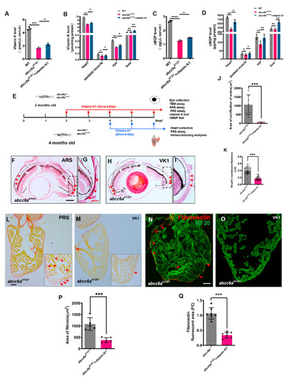
Vitamin K treatment relives ocular calcification and cardiac fibrosis in abcc6aΔ1/Δ1 mutants. (A–D) Graphical representation of sandwich ELISA measurements for tissue and serum vitamin K and cMGP levels in WT (n = 18) and DMSO/ethanol- (n = 18) or vitamin K1-treated abcc6aΔ1/Δ1 fish (n = 18) fish. * p < 0.05, ** p < 0.01, *** p < 0.001, **** p < 0.0001, Student’s t-test (unpaired, two-tailed). (E) Experimental design for once-a-day DMSO/ethanol (0.1%) and vitamin K1 (80 µM) treatment of abcc6a+/+ and abcc6aΔ1/Δ1 fish. Red arrows indicate experimental steps, changing water every day for 4 months from 2 months old; for eye collection, TEM, ARS, and PRS assay, vitamin K and cMGP tests. Blue arrows indicate experimental steps for heart collection, PRS assay and immunostaining analyses for 2 months from 4 months of age. (F–I) Vitamin K rescues abnormal scleral calcification in abcc6aΔ1/Δ1 mutant zebrafish. (G,I) Higher-magnification images of the dashed boxes in (F,H). Dashed black line indicates approximate plane of resection. Red arrow marks abnormal calcification of scleral layer in the abcc6aΔ1/Δ1 mutant eyes. (J) Quantification of area of calcification of sclera after 4 months of DMSO/ethanol (n = 7) or vitamin K1 treated treatment in abcc6aΔ1/Δ1 fish (n = 7). *** p < 0.001, Student’s t-test (unpaired, two-tailed). (K) Quantification of Bruch’s membrane thickness after 4 months of DMSO/ethanol (n = 13, 8 measuring points per sample) or vitamin K1 treatment in abcc6aΔ1/Δ1 fish (n = 15, 8 measuring points per sample). *** p < 0.001, Student’s t-test (unpaired, two-tailed). (L,M) Picrosirius Red staining of heart sections shows significantly reduced collagen deposition reside in the compact layer and trabeculae after treatment with vitamin K1 (M), as compared to abcc6aΔ1/Δ1 mutant hearts after treatment with DMSO/ethanol (L). Red for collagen (L; Red arrow) and yellow for cardiomyocyte fiber. (N,O) Immunofluorescent section images of adult ventricles stained with anti-Fibronectin and anti-MF20 antibodies from DMSO/ethanol-treated (N) or vitamin K1 treated abcc6aΔ1/Δ1 mutant fish (O), showing that vitamin K reduces Fibronectin. (P) Quantification of area of fibrosis after 2 months of DMSO/ethanol (n = 5) or vitamin K1 treatment in abcc6aΔ1/Δ1 fish (n = 5). *** p < 0.001, Student’s t-test (unpaired, two-tailed). (Q) Quantification of Fibronectin fluorescent area after 2 months of DMSO/ethanol (n = 7) or vitamin K1 treatment in abcc6aΔ1/Δ1 fish (n = 7). *** p < 0.001, Student’s t-test (unpaired, two-tailed). Scale bar: 250 μm (F,H); 50 μm (L–O); 125 μm (G,I).
|
