|
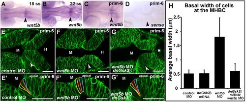
Wnt5b regulates basal constriction possibly through Gsk3β. (A–D) In situ hybridization of wnt5b expression during MHB development at 18 ss (A), 22 ss (B), and prim-6 (C). (D) prim-6 sense probe control. (E–G′) Live confocal images of the MHB region in prim-6 stage embryos. Single-cell wild-type embryos were injected with mGFP to label cell membranes and co-injected with control MO (E,E′), wnt5b MO (F,F′), or wnt5b MO and dnGsk3β mRNA (G,G′). (H) Quantification of basal cell width in control MO, wnt5b MO, dnGsk3β mRNA (image not shown), and wnt5b MO+dnGsk3β mRNA injected embryos. (H) For each treatment group, n=3 embryos. For each embryo, 6 cells located at the MHBC were measured, 3 cells on each side. Arrowheads indicate MHBC. M, midbrain; H, hindbrain. Scale bars: 26 µm.
|