
Fig. S2
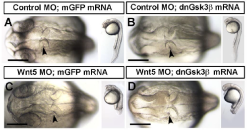
Gross morphology images of dnGsk3 and wild-type Gsk3 overexpression phenotypes and dnGsk3 rescue of wnt5b morphants. (A-D) Brightfield dorsal and lateral images of control MO (A,B) and wnt5b MO (C,D) injected embryos, co-injected with mGFP (A,C), or dnGsk3 (B,D) mRNA. (A) Control morphants co-injected with mGFP mRNA demonstrating a normal MHBC basal constriction phenotype. (B) Control morphants co-injected with dnGsk3 mRNA exhibit an eyeless phenotype, but undergo basal constriction normally with this concentration of dnGsk3 (n=9). (C) Wnt5b morphants co-injected with control mRNA exhibit abnormal MHBC morphogenesis, tail defects, and fail to undergo basal constriction (n=6). (D) Wnt5b morphants co-injected with dnGsk3 mRNA exhibit a loss of eyes and tail defects, but the gross morphology of basal constriction is rescued and occurs normally (n=6). Arrowheads indicate the MHBC. Scale bars: 100m. |