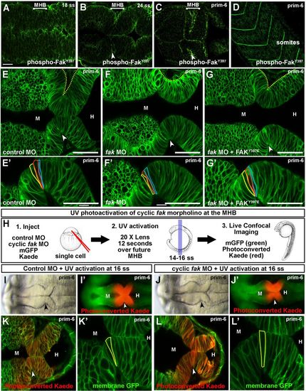
Fak is required at the MHB for basal constriction. (A–D) Wild-type embryos stained with anti-phospho-FakY397 antibody and imaged by scanning confocal microscopy. (A,B) phospho-FakY397 is localized at the basal and apical sides of the neural tube at 18 and 24 ss. (C) Activated Fak is enriched at the MHBC at prim-6. (D) Phospho-FakY397 is localized to somite boundaries at prim-6. (E–G′) Live confocal images of embryos co-injected with mGFP and control MO (E,E′), fak MO (F,F′), or fak MO+FAKY397E mRNA (G,G′). (E′–G′) Magnifications of individual cells outlined at the MHBC. (H) Schematic for fak caged MO experiments. (1.) One-cell stage wild-type embryos were co-injected with mGFP and photoconvertable Kaede mRNA and either control MO or cyclic fak MO. (2.) Cyclic fak MO was uncaged at 16 ss by UV activation. (3.) Embryos were incubated until prim-6 and then imaged using brightfield, fluorescence, and live scanning confocal microscopy. (I–J′) Gross morphology using brightfield imaging (I,J) and corresponding fluorescent (I′,J′) images of embryos injected with control MO (I,I′) or photoactivatable fak MO (J,J′) after UV photoconversion. (K–L′) Live confocal images showing the MHB region of prim-6 embryos after photoconversion. (K′,L′) Magnifications of the neuroepithelium shown in K and L with individual cells outlined at the MHBC. (n>6). Anterior is to the left in all images. Arrowheads indicate MHBC. M, midbrain; H, hindbrain. Scale bars: A–C=20 µm. E–G′=50 µm.
|
