|
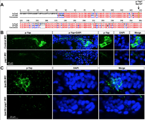
Validation of the p-‐Yap1 antibody used in the study. (A) Alignment of the N-‐terminal amino acid sequences of human (hsYAP1) and zebrafish Yap1 (drYap1) proteins. High concensus sequences (>90%) are shown in red, and low concensus sequences (>50%) are depicted in blue. Arrow, the Ser127 phosphorylation site recognized by the antibody. (B)Confocal images of the pronephric duct region stained with p-‐Yap1 and DAPI in 32 hpf embryo injected with control-‐ of yap1 MO. Images in the left 2 panels are generated from maximum projection of 6 confocal z-‐stacks. A single z-‐stack of the boxed areas is depicted in the right 3 panels. Note the p-‐Yap1 signal is in the cytoplasm and does not overlap with the nucleus. (C) Confocal images of the pLLP cells stained with p-‐Yap1 and DAPI in embryos injected with indicated MOs. The p-‐Yap1 signal is in the cytosol of pLLP cells.
|