
Fig. S14
- ID
- ZDB-FIG-180529-13
- Publication
- Sánchez-Iranzo et al., 2018 - Tbx5a lineage tracing shows cardiomyocyte plasticity during zebrafish heart regeneration
- Other Figures
- All Figure Page
- Back to All Figure Page
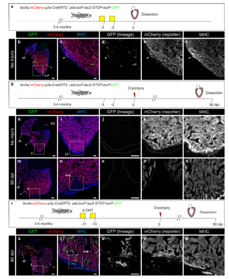
Trabecular cardiomyocyte lineage tracing control experiments. a–f tbx5a:mCherryp-2a-CreERT2 was crossed into ubb:loxP-lacZ-STOPloxP- GFP. Fish were treated with 4-Hydroxytamoxifen (4-OHT) at days 3 and 2 before fixation. Arrowhead points to one of the 2 cells GFP+ cells identified in 1 out of 6 hearts. g– q loxP sites in tbx5a:Cherryp-p2a-CreERT2;ubb:loxP-lacZ-STOP-loxP-GFP do not recombine in the absence of 4-OHT. g tbx5a:Cherry-p2A-CreERT2 was crossed into ubb:loxP-lacZ-STOP-loxP-GFP. Fish were cryoinjured but not treated with 4-OHT. h–l Heart before injury. No recombined cells were observed (n = 4/4). m–q Hearts were injured and dissected at 90 days postinjury (dpi). No GFP+ recombined cells were visible at the regenerated area (n= 3/3). r–w Recombination is not due to the persistence of 4-OHT in the adult fish beyond cryoinjury. r tbx5a:mCherry-p2a-CreERT2 was crossed into ubb:loxPlacZ- STOP-loxP-GFP. Fish were treated with 4-OHT during days 11 to 10 before the injury. s–w Contribution of the GFP+ trabeculae to the new compact layer and tbx5a:mCherry switch off in these cells can be observed (n=3/3). at, atrium; v, ventricle. Scale bars, b, h, m 100 μm, c, i, j, n, o, t, u 25 μm |