
Fig. 2
- ID
- ZDB-FIG-180524-14
- Publication
- Sánchez-Iranzo et al., 2018 - Tbx5a lineage tracing shows cardiomyocyte plasticity during zebrafish heart regeneration
- Other Figures
- All Figure Page
- Back to All Figure Page
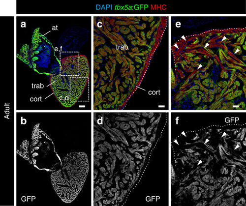
Expression profile of tbx5a-positive cardiomyocytes in adult zebrafish hearts. a, b Sagittal sections through tbx5a:GFP adult uninjured heart immunostained with GFP (green) and Myosin Heavy Chain (MHC; red). Nuclei are counterstained with DAPI (blue). c–e Zoomed views of boxed area in a. b, d, f Single channels for GFP. The trabecular myocardium is tbx5a:GFP+, whereas the cortical layer is tbx5a:GFP−. Note tbx5a:GFP− cardiomyocytes (arrowheads) in the basal part of the ventricle close to the atrioventricular canal (n = 13/13). at, atrium; cort, cortical layer; trab, trabecular layer. Scale bars, a, b 100 μm and c–f 25 μm |