
Fig. S13
- ID
- ZDB-FIG-180529-11
- Publication
- Sánchez-Iranzo et al., 2018 - Tbx5a lineage tracing shows cardiomyocyte plasticity during zebrafish heart regeneration
- Other Figures
- All Figure Page
- Back to All Figure Page
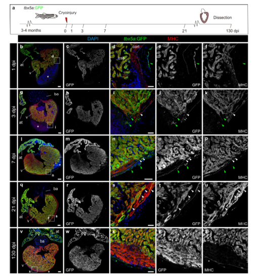
Expression of tbx5a:GFP during adult heart regeneration. a, Illustration of the experimental setup. b–z Sagittal sections through tbx5a:GFP hearts at 1 (b–f n=4/4), 3 (g–k n=4/5), 7 (l–p n=3/3), 21 (q–u n=4/4) and 130 (v–z n=4/4) days post injury (dpi). Sections were immunostained with anti-GFP (green) and anti-Myosin Heavy Chain (MHC; red). Nuclei are counterstained with DAPI (blue). Asterisks indicates the injury area, dotted line marks the outer border of the cortical layer. tbx5a:GFP expression is limited to the trabecular myocardium. Few myosin-negative tbx5a:GFP+ cells are found within the epicardial layer (arrowhead). At 130 dpi, a tbx5a:GFP- thickened cortical myocardium covers a tbx5a:GFP+ trabecular myocardium at the injury site (v–z). White arrowheads point to tbx5a:GFP+ cardiomyocytes; green arrowheads label tbx5a:GFP+ noncardiomyocytes. Scale bars, b, g, l, q, v 100 μm, d, i, n, s, x 25 μm |