
Fig. 6
- ID
- ZDB-FIG-180524-18
- Publication
- Sánchez-Iranzo et al., 2018 - Tbx5a lineage tracing shows cardiomyocyte plasticity during zebrafish heart regeneration
- Other Figures
- All Figure Page
- Back to All Figure Page
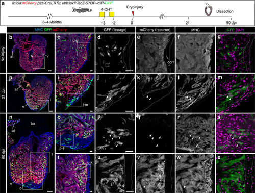
Contribution of tbx5a-derived cells during regeneration of the adult zebrafish heart. a tbx5a:Cherry-p2A-CreERT2 was crossed into ubb:loxP-lacZ-STOP-loxP-GFP. 4-Hydroxytamoxifen (4-OHT) was added 2 and 3 days before cryoinjury, to induce recombination of loxP sites. Hearts were fixed at 21 and 90 days postinjury (dpi) and sectioned for immunofluorescent detection of GFP+ tbx5a-derived cells and mCherry+ tbx5a-expressing cells. Nuclei were counterstained with DAPI. b In the uninjured heart (n = 7/7), mCherry expression was homogeneous in the trabecular myocardium and absent in the cortical layer. GFP+ cells were found in the trabecular layer. c Zoomed view of boxed area in b. d–f Single channels of boxed area shown in c. g GFP and DAPI channels only. h, n Section of hearts at 21 dpi h and 90 dpi n. Upon cryoinjury to the ventricular apex, tbx5a-derived cardiomyocytes were present also in the cortical layer, particularly at the site of injury (h–m, n = 6/7; o–s, n = 5/6; t–x, n = 6/6), whereas tbx5a+ cardiomyocytes in general were restricted to the trabecular myocardium (n = 5/6) t–x. Nuclear counterstaining revealed GFP+ cell bodies in the cortical layer (arrowheads). at, atrium; cort, cortical layer; ba, bulbus arteriosus; v, ventricle. Scale bars, b, h,n 100 µm and c, d, i, j, o, p, t, u 25 μm |