
Fig. S1
- ID
- ZDB-FIG-180524-23
- Publication
- Sánchez-Iranzo et al., 2018 - Tbx5a lineage tracing shows cardiomyocyte plasticity during zebrafish heart regeneration
- Other Figures
- All Figure Page
- Back to All Figure Page
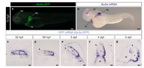
Characterization of the tbx5a:GFP reporter line. a Lateral view of a tbx5a:GFP larvae at 3 days postfertilisation. A merged fluorescent and brightfield image is shown. b mRNA in situ hybridization (ISH) with a tbx5a antisense riboprobe on the same staged larvae as the one shown in a (n=7/7). c–g gfp mRNA on embryonic heart sections of tbx5a:GFP fish at different stages of development. The black arrowheads point to the tbx5a:GFP- area of the ventricle. at, atrium; dpf, days postfertilisation; hpf, hours postfertilisation; ht, heart tube; ret, retina; v, ventricle. Scale bars, a, b 100 μm and c, g 25 μm |