Fig. 2
- ID
- ZDB-FIG-170922-49
- Publication
- Xue et al., 2017 - The Vascular Niche Regulates Hematopoietic Stem and Progenitor Cell Lodgment and Expansion via klf6a-ccl25b
- Other Figures
- All Figure Page
- Back to All Figure Page
|
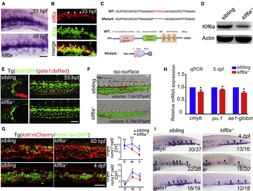
The klf6a-Deficient Embryos Display HSPC Defects in the CHT (A) The high-resolution expression pattern of klf6a in the CHT at 33 hpf and 48 hpf. Black arrowheads indicate the expression of klf6a in caudal venous ECs. Scale bar, 30 μm. (B) Double FISH shows klf6a (red) co-localized with fli1a (green) at 33 hpf in the CHT. White arrowheads indicate the expression of klf6a, fli1a or the co-expression of fli1a and klf6a, respectively. Scale bar, 30 μm. (C) The mutation of klf6a with a 7-bp deletion in genomic DNA and the predicated truncated protein. (D) Western blot result of Klf6a in the siblings and mutants at 48 hpf. (E) Confocal imaging showing kdrl+ vascular deficiency in the CHT and head region, whereas gata1+ cells accumulated in the brain in klf6a−/− embryos in Tg (kdrl:GFP/gata1:dsRed) background. Scale bar, 50 μm. (F) 3D imaging of the CHT vascular structure. Scale bar, 50 μm. (G) Confocal imaging of runx1+ cells and vascular structure in the siblings and klf6a−/− embryos in Tg (kdrl:mCherrry/runx1:en-GPF) background at 60 hpf and 4 dpf (mean ± SD, t test; ∗p < 0.05, ns [not significant] > 0.05, n = 5). Scale bar, 50 μm. (H) The qPCR results of cmyb, pu.1, and ae1-globin expression in siblings and klf6a−/− embryos at 5 dpf (mean ± SD, t test; ∗p < 0.05, n = 3). (I) Expression of cmyb, lyz, and gata1 in siblings and klf6a−/− embryos at 4 dpf by WISH. Black arrowheads indicate the expression pattern of these markers. See also Figure S2. |
| Genes: | |
|---|---|
| Antibody: | |
| Fish: | |
| Anatomical Terms: | |
| Stage Range: | Prim-15 to Day 5 |
| Fish: | |
|---|---|
| Observed In: | |
| Stage Range: | Long-pec to Day 5 |
Reprinted from Developmental Cell, 42(4), Xue, Y., Lv, J., Zhang, C., Wang, L., Ma, D., Liu, F., The Vascular Niche Regulates Hematopoietic Stem and Progenitor Cell Lodgment and Expansion via klf6a-ccl25b, 349-362.e4, Copyright (2017) with permission from Elsevier. Full text @ Dev. Cell

