
Fig. 5
- ID
- ZDB-FIG-160525-38
- Publication
- San et al., 2016 - Normal formation of a vertebrate body plan and loss of tissue maintenance in the absence of ezh2
- Other Figures
- All Figure Page
- Back to All Figure Page
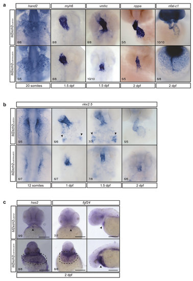
Myocardial development is affected in MZezh2 embryos. (a) In situ hybridization for different heart markers in MZezh2hu5670/+ and MZezh2hu5670/hu5670 at various time points of development. hand2 is an early myocardial marker. myh6 is a marker for atrial cells. vmhc is a marker for ventricular cells. nppa is a late myocardial marker. nfat-c1 is an endocardial marker. All these markers are expressed in MZezh2hu5670/hu5670, although vmhc, nppa, and nfat-c1 expression show a smaller number of positive cells. (b) In situ hybridization for nkx2.5 at different time points after fertilization in MZezh2hu5670/+ and MZezh2hu5670/hu5670 embryos. Arrow heads point to cells of the pharyngeal arch artery progenitors. This is absent in MZezh2hu5670/hu5670. (c) In situ hybridization for has2 and fgf24 at 2 dpf in MZezh2hu5670/hu5670 and their heterozygous siblings. In MZezh2hu5670/+ expression is restricted to the heart (arrow heads), whereas in the MZezh2hu5670/hu5670 embryos expression is visible in the area surrounding the heart tube (encircled by dashed line). For fgf24 this is also shown from a lateral view (arrow heads). Scale bar is 200 µm. The numbers indicate the number of embryos with the displayed phenotype compared to the total number of embryos analyzed. |
| Genes: | |
|---|---|
| Fish: | |
| Anatomical Terms: | |
| Stage Range: | 10-13 somites to Long-pec |
| Fish: | |
|---|---|
| Observed In: | |
| Stage Range: | Prim-5 to Long-pec |