
Fig. 6
- ID
- ZDB-FIG-160316-26
- Publication
- Xu et al., 2016 - Four and a Half LIM Domains 1b (Fhl1b) Is Essential for Regulating the Liver versus Pancreas Fate Decision and for β-Cell Regeneration
- Other Figures
- All Figure Page
- Back to All Figure Page
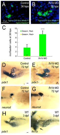
Reduction of Fhl1b activity enhances the capacity of β-cell regeneration. (A and B) Confocal images of [Tg(ins:CFP-NTR)s892; Tg(ins:Kaede)jh6] control larvae (A) and fhl1b MO-injected (B) larvae at 36 hours-post-ablation (hpa) stained with Topro (blue). A greater number of β-cells regenerated in recovering fhl1b-MO injected larvae (B) compared to that of control larvae (A). (C) Quantification of the number (mean±SD) of regenerated and survived β-cells. After photoconversion and ablation, the survived β-cells are red and green (yielding a combined color of yellow), whereas the newly formed β-cells are green only. 3.8±1.3 β-cells were green-only-positive in control recovering larvae, while 9.6±1.4 β-cells expressed as green-only in fhl1b-MO injected recovering larvae. Almost no β-cells survived the ablation in both the control and fhl1b-MO injected recovering larvae. Cells in 20 planes of confocal images from 10 individual larvae were counted. Asterisks indicate statistical significance: ***, P < 0.001. (D-G) Whole-mount in situ hybridization showing the expression of pdx1 (D and E) and neurod (F and G) at 72 hpf, comparing control embryos (D and F) and fhl1b morphants (E and G). pdx1 is expressed in the pancreas including the principal islet (white dotted circles), the HPD system (white brackets), and the proximal intestine, but not in the liver. neurod is expressed mainly in the principal islet (white dotted circles) with slight expression in the HPD system (white brackets). pdx1 (E) and neurod (G) expression in the principal islet and the HPD system was greatly increased in fhl1b morphants. (H-I) Whole-mount in situ hybridization showing the expression of pdx1 (H) and fhl1b (I) in wild-type embryos at 3 dpf. (H) pdx1 is expressed in the pancreas including the principal islet, the HPD system (white bracket), and the proximal intestine (white dotted line), but not in the liver. (I) fhl1b is expressed at high levels in the liver cells (black arrow), which never express pdx1, whereas the HPD system (white bracket) expresses low levels of fhl1b. Most pancreatic cells except for a few cells in the principal islet (yellow arrow) do not express fhl1b. The distal intestine also expresses fhl1b (white dotted lines). A-B, confocal projection images, ventral views, anterior to the top. D-I, dorsal views, anterior to the left. Scale bars, 20 µm. |
| Genes: | |
|---|---|
| Fish: | |
| Condition: | |
| Knockdown Reagents: | |
| Anatomical Terms: | |
| Stage Range: | Protruding-mouth to Day 6 |
| Fish: | |
|---|---|
| Condition: | |
| Knockdown Reagents: | |
| Observed In: | |
| Stage Range: | Protruding-mouth to Day 6 |