
Fig. 2
- ID
- ZDB-FIG-160316-22
- Publication
- Xu et al., 2016 - Four and a Half LIM Domains 1b (Fhl1b) Is Essential for Regulating the Liver versus Pancreas Fate Decision and for β-Cell Regeneration
- Other Figures
- All Figure Page
- Back to All Figure Page
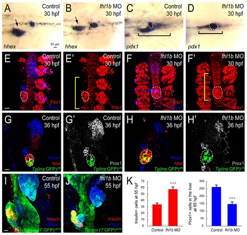
Loss of Fhl1b activity enhances induction of pancreatic cells and compromises liver specification. (A-D) Whole-mount in situ hybridization showing the expression of hhex (A and B) and pdx1 (C and D), comparing control embryos (A and C) and fhl1b morphants (B and D) at 30 hpf. hhex is expressed in the liver (black arrows) and the dorsal pancreatic bud (white dotted circles). pdx1 is expressed in the developing pancreas including the dorsal pancreatic bud (white dotted circles) and intestine (black brackets), but not in the liver. hhex expression was reduced in the liver of fhl1b morphants, while expanded in the dorsal pancreatic bud (B). pdx1 expression in the dorsal pancreatic bud was also expanded, while its expression in the intestinal bulb primordium appeared to be reduced in fhl1b morphants (D) compared to that of control embryos (C). (E–F′) Confocal images of control embryos (E and E′) and fhl1b morphants (F and F′) at 30 hpf, stained for Pdx1 (red; expression in the dorsal pancreatic bud is outlined by white dotted circles) and Prox1 (blue). The somites are also Pdx1 positive. Compared to that of control embryos (E and E′), in fhl1b morphants (F and F′), the Pdx1 expression domain in the dorsal pancreatic bud was expanded, while the Prox1 expression domain was reduced. Note that the Pdx1 expression domain in the intestinal primordium (yellow brackets) appeared to be decreased in morphants (F′). (G-H′) Confocal images of Tg(ins:GFP)zf5 control embryos (G and G′) and fhl1b morphants (H and H′) at 36 hpf, stained for Islet (red; expression in the dorsal pancreatic bud is outlined by white dotted circles) and Prox1 (blue in G and H; grey in G′ and H′). In fhl1b morphants (H and H′), the Prox1 expression domain was greatly reduced, whereas the number of Tg(ins:GFP)zf5- and Islet-expressing cells was increased. (I and J) Confocal images of Tg(sox17:GFP)s870 control embryos (I) and fhl1b morphants (J) at 55 hpf, stained for Insulin (red; outlined by white dotted circles) and Prox1 (blue). fhl1b morphants (J) continuously exhibited an enlarged Insulin-expressing β-cell population with a reduced number of Prox1-positive cells in the liver. (K) Quantification of the number (mean±SD) of Insulin-positive cells in the pancreas (red) and Prox1-positive cells in the liver (blue) at 55 hpf. 33.9±2.1 cells were Insulin-positive in control embryos, whereas 57.8±3.6 cells were Insulin-positive in fhl1b morphants. 148.0±15.2 cells expressed Prox1 in fhl1b morphants, while 262.7±14.0 cells were Prox1-positive in control embryos. Cells in 20 planes of confocal images from 5 individual embryos were counted. Asterisks indicate statistical significance: ***, P < 0.001. A-D, dorsal views, anterior to the left. E-J, confocal projection images, ventral views, anterior to the top. Scale bars, 20 µm. |
| Genes: | |
|---|---|
| Antibody: | |
| Fish: | |
| Knockdown Reagents: | |
| Anatomical Terms: | |
| Stage Range: | Prim-15 to Long-pec |
| Fish: | |
|---|---|
| Knockdown Reagents: | |
| Observed In: | |
| Stage Range: | Prim-15 to Long-pec |