|
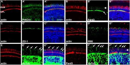
Spatial distribution of cell junction proteins and cilia in 5 dpf zebrafish. (A–E) Left-most panels show transverse cryosections stained with phalloidin (red) to label actin in the adherens junctions at the outer limiting membrane (OLM) and the outer plexiform layer (OPL). The middle panels show staining for determinants of apico-basal polarity (green), while the right-most panels show the merged images. (A1–A3) PrkCi/z immunoreactivity. (B1–B3) Pard3 immunoreactivity localizes to the OPL (arrow) and at cell junctions (arrowhead). (C1–C3) ZO-1 staining at the OLM. (D1–D3) Ift88 immunoreactivity stains cilia. (E1–E3) Acetylated tubulin immunoreactivity was seen in cilia and neural processes throughout the inner nuclear layer and plexiform layers. (F1–F3) Pard3 staining (red) was seen at the OLM (arrowhead), but did not colocalize with acetylated tubulin signal in the cilia (arrows). All sections were counterstained with DAPI (blue). Scale bar = 10 μm.
|