
Fig. 2
- ID
- ZDB-FIG-150316-4
- Publication
- Maier et al., 2014 - RA and FGF Signalling Are Required in the Zebrafish Otic Vesicle to Pattern and Maintain Ventral Otic Identities
- Other Figures
- All Figure Page
- Back to All Figure Page
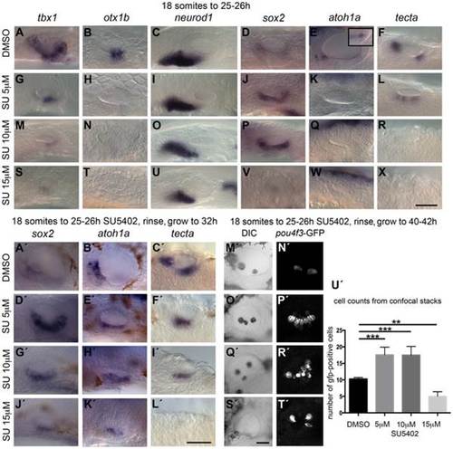
Inhibition of FGF signalling by SU5402 reveals a concentration-dependent effect of FGF signalling during OV development. Wild-type (WT) embryos were treated with DMSO or SU5402 from 18S to 26 hpf (A–X) or rinsed and grown on to 32 hpf (A2–L2) or 40–42 hpf (M2–T2). (A–F) WT embryos treated with DMSO show normal expression of non-neural markers tbx1 (n = 51) and otx1b (n = 61; A,B), the neuronal marker neurod1 (n = 64; C), and the sensory markers sox2 (n = 56), atoh1a (n = 26) and tecta (n = 44; D–F). Boxed area in panel (E) depicts the atoh1a expression in posterior sensory patch in a different focal plane. (G–R) Treatment with 5 µM SU5402 (G–L) or 10 µM SU5402 (M–R) reduces the expression of tbx1 (n = 56; G,I) and neurod1 (n = 76; M,O). Expression of otx1b is lost (n = 32; H,N) and expression of sox2 is extended in the ventral OV floor (n = 45; J,P). The expression of atoh1a is lost (n = 18; K,Q) and expression of tecta is weak but extended (n = 27; L, 5 µM SU5402) or lost (n = 40; R, 10 µM SU5402). (S–X) Treatment with 15 µM SU5402 reduces the expression of tbx1 (n = 38; S) and neurod1 (n = 65; U); expression of otx1b (n = 57), sox2 (n = 58), atoh1a (n = 35), and tecta (n = 51) is lost (T,V–X). (A2–C2) WT embryos treated with DMSO show normal expression of the sensory markers sox2 (n = 23), atoh1a (n = 31) and tecta (n = 25) at 32 hpf. (D2–I2) In embryos treated with 5 µM SU5402 (D2–F2) or 10 µM SU5402 (G2–I2), expression of sox2 (n = 23) and atoh1a (n = 25) is extended in the ventral OV floor; tecta expression is misplaced in a single ventromedial patch (n = 24). (J2–L2) Treatment with 15 µM SU5402 reduces or abolishes the expression of sox2 (n = 18), atoh1a (n = 16), and tecta (n = 14). (M2,N2) Tg(pou4f3:GFP) embryos treated with DMSO show normal hair cell patterns marked with GFP (n = 26). (O2,P2) In Tg(pou4f3:GFP) embryos treated with 5 µM SU5402, GFP-positive cells extended between the anterior and posterior sensory patch in the ventral OV floor (n = 18). (Q2,R2) In Tg(pou4f3:GFP) embryos treated with 10 µM SU5402, GFP-positive cells appear less orderly and are extended in the ventral OV floor (n = 12). (S2,T2) Treatment with 15 µM SU5402 reduced the number of GFP-positive cells in the OV (n = 14). (U2) Graphical representation of counts of GFP-positive cells at 40–42 hpf. Error bars represent standard error of the mean. One-way ANOVA with Dunnett′s multiple comparison post-test: **p = 0.0065, ***p<0.0005. All panels are lateral views with anterior to the left, apart from the panels depicting tecta (dorsal views; anterior to the left, lateral up). Scale bar: 50 µm. |
| Genes: | |
|---|---|
| Fish: | |
| Condition: | |
| Anatomical Terms: | |
| Stage Range: | Prim-5 to Prim-25 |
| Fish: | |
|---|---|
| Condition: | |
| Observed In: | |
| Stage: | Prim-25 |