
Fig. 7
- ID
- ZDB-FIG-140714-42
- Publication
- van Ham et al., 2014 - Intravital correlated microscopy reveals differential macrophage and microglial dynamics during resolution of neuroinflammation
- Other Figures
- All Figure Page
- Back to All Figure Page
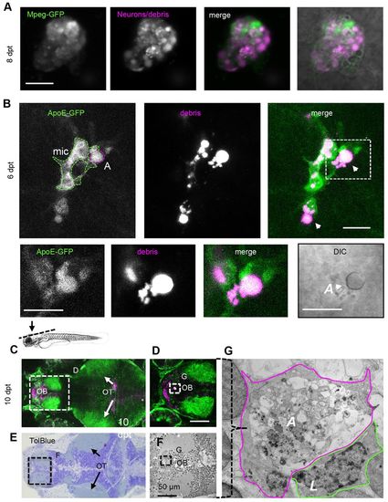
Apoptotic phagocytes are engulfed by microglia. (A) Large mpeg-GFP-expressing phagocyte 8 days post-treatment (dpt) showing cytoplasmic mCherry-positive vacuoles (magenta) and mpeg-GFP expression (green), suggestive of an apoptotic phagocyte. (B) ApoE-expressing microglia (mic; green) phagocytizing non-motile, apoptotic phagocytea (‘A’; magenta, arrowheads) (supplementary material Movie 9). The boxed area is shown below at higher magnification. The DIC image at the bottom right shows that the phagocyte being engulfed by microglia is apoptotic (reflective button-like rounded cell body). Data were recorded using intravital imaging of three distinct channels to visualize NTR, ApoE-GFP and apoptotic cells. The schematic above C indicates the region shown in C and E. (C) Optical section of live forebrain and optic tectum region of NTR animals treated with MTZ 10 days after treatment; green indicates GFP expression (supplementary material Movie 10). (D) Higher magnification of forebrain as shown in C. (E) Toluidine Blue stained section of region of brain of same animal as shown in C. (F) Electron micrograph of region indicated in E. (G) Electron micrograph of phagocytic profile observed in live imaging, showing large apoptotic cytoplasm including vacuoles and lysosomal debris demarcated with solid purple line, partly surrounded by a leukocytic cell (‘L’). ‘A’, apoptotic phagocyte; mic, microglia; OB, olfactory bulb; OT, optic tecti. Scale bars: 10 μm (A,B), 50 μm (D). See also supplementary material Movies 9–11. |