
Fig. 3
- ID
- ZDB-FIG-140714-38
- Publication
- van Ham et al., 2014 - Intravital correlated microscopy reveals differential macrophage and microglial dynamics during resolution of neuroinflammation
- Other Figures
- All Figure Page
- Back to All Figure Page
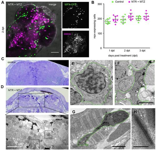
Neutrophils are not affected by cell ablation in the brain. (A) Myeloperoxidase (mpx)-expressing neutrophils (green) in NTR MTZ-treated brain (magenta) 2 days post-treatment. (B) Total numbers of mpx-expressing neutrophils in whole animal controls and 1–3 days post-treatment (n=10 animals). Numbers do not differ significantly. (C,D) Toluidine Blue stained 1 micron section of brain from control and NTR MTZ-treated animals. Control brain show homogeneous cellular profiles, whereas NTR animals treated with MTZ shows cells irregular in staining density, cytoplasmic inclusions and dark pyknotic nuclei. (D, lower panel) Electron micrograph of region marked in D shows features not found in control brains, including dark stained cells, phagocytic leukocytes and spongy appearance of tissue (also see Fig. 4). (E) High magnification of monocyte-like cell in D. (F) High magnification view of phagocytic cell in D. (G,H) Neutrophil (G) marked by cigar-shaped granules (H), located between jaw muscle cells, showing characteristic striping pattern not found in the brain. Scale bars: 50 μm (A,D, lower panel), 5 μm (F,G), 200 nm (H). |