
Fig. 5
- ID
- ZDB-FIG-120710-48
- Publication
- Choorapoikayil et al., 2012 - Analysis of her1 and her7 Mutants Reveals a Spatio Temporal Separation of the Somite Clock Module
- Other Figures
- All Figure Page
- Back to All Figure Page
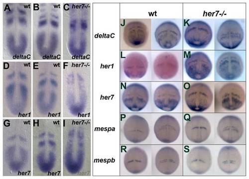
Expression analysis of segmentation genes in her7hu2526 mutant embryos. In situ hybridisation analysis of deltaC, her1 and her7 in wild type (A, B and D, E and G, H, respectively) and her7hu2526 mutants (C, F, I, respectively) at 10–12 somite stage and between 90% epiboly and bud stage (J, L, N for wild type expression patterns and K, M, O for respective expression patterns in the mutant embryos). Expression patterns of deltaC, her1 and her7 at 10–12 somite stage are disrupted in the mutant appear unperturbed between 90% epiboly and bud stage. Expression patterns of mespa and mespb are not affected in the her7hu2526 mutant between 90% epiboly and bud stage (Q and S, respectively) and similar to the wild type (P and R, respectively). |
| Genes: | |
|---|---|
| Fish: | |
| Anatomical Terms: | |
| Stage Range: | 90%-epiboly to 10-13 somites |