|
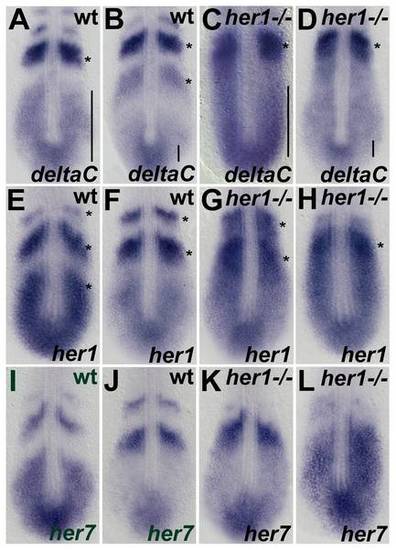
Expression analysis of the segmentation clock genes at 10–12 somite stage in her1hu2124 mutants. In situ hybridisation analysis of the segmentation clock genes deltaC, her1, and her7, in wild type embryos (A,B,E,F,I,J) her1hu2124 mutant (C,D,G,H,K,L) at the 10–12 somite stage. Two significantly different patterns are shown for each gene to indicate oscillatory expression. Expression of deltaC in her1hu2124 mutants at this developmental stage is identical to the 90% epiboly (see Fig. 2D), cyclic in the posterior PSM and disrupted expression in the anterior PSM (C, D) compared to wild type (A, B). Expression of her1 and her7 oscillates in the her1hu2124 mutant but on average one expression stripe is lacking (see asterisks in G, H and K, L, respectively) compared to the respective wild type expression domains (asterisks in E, F and I, J). Further, the patterns in the PSM of mutants appear stretched towards the anterior compared to wild type (see bars in A-D) suggesting that one expression wave is lacking. Dorsal view, anterior to the top.
|