
Fig. 2
- ID
- ZDB-FIG-120710-45
- Publication
- Choorapoikayil et al., 2012 - Analysis of her1 and her7 Mutants Reveals a Spatio Temporal Separation of the Somite Clock Module
- Other Figures
- All Figure Page
- Back to All Figure Page
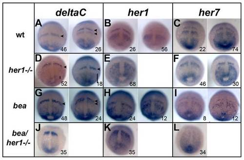
Expression analysis of segmentation genes in her1 hu212 and beatm98 mutants. In situ hybridisation analysis of segmentation clock genes deltaC, her1, and her7 in wild type (A–C) her1hu2124 mutants (D–F), bea tm98 mutants (G–I) and her1hu2124/bea tm98 double mutants (J–L) at 90% epiboly. Cyclic deltaC expression is disrupted in the anterior PSM of her1hu2124 mutants. Instead of one or two expression stripes as in the wild type (A, arrowheads) only one stripe of expression is observed (D, arrowhead). Expression domains in the posterior PSM display different sizes indicating unperturbed oscillation of deltaC in the tail bud of her1hu2124 mutants (D, bars). Cyclic expression of her1 is fully disrupted in the her1hu2124 mutant (E) when compared to wild type (B), whereas her7 expression remains oscillatory (compare C and F). Cyclic expression of all three genes is observed in beatm98 although some slight initial perturbation is observed (G-I). In her1hu2124/beatm98 double mutants, all three clock genes show fully disrupted expression patterns at 90% epiboly. Dorsal views, anterior to the top, number in each panel indicate cycling phases. |
| Genes: | |
|---|---|
| Fish: | |
| Anatomical Terms: | |
| Stage Range: | 90%-epiboly to Bud |