
Fig. 3
- ID
- ZDB-FIG-111025-45
- Publication
- Sun et al., 2007 - Epibranchial and otic placodes are induced by a common Fgf signal, but their subsequent development is independent
- Other Figures
- All Figure Page
- Back to All Figure Page
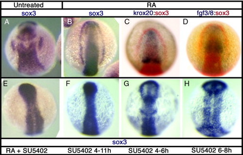
sox3 expression is affected by disruption of Fgf signaling. In situ hybridization for sox3 expression at 11 hpf in DMSO treated control embryos (A) or following treatments to alter Fgf signaling. Treatment with retinoic acid (RA) from 6–7 hpf causes expansion of the hindbrain, as shown by rostral displacement of the rhombomere 3 band of krox20 expression (C) and consequently expression of fgf3/8 throughout most of the rostral CNS (D). This treatment results in a striking expansion of sox3 expression, in the most severe cases resulting in a complete arc of expression around the anterior of the embryo (B). Expression of sox3 outside of the CNS, even in the presence of RA, is completely blocked when Fgf signaling is inhibited using the small molecule inhibitor, SU5402 (E–H). SU5402 causes a loss of placodal sox3 expression when treated from 4–11 hpf (F), but also after shorter treatments at 4–6 hpf (G) and 6–8 hpf (H). |
| Genes: | |
|---|---|
| Fish: | |
| Conditions: | |
| Anatomical Terms: | |
| Stage: | 1-4 somites |
| Fish: | |
|---|---|
| Conditions: | |
| Observed In: | |
| Stage: | 1-4 somites |
Reprinted from Developmental Biology, 303(2), Sun, S.K., Dee, C.T., Tripathi, V.B., Rengifo, A., Hirst, C.S., and Scotting, P.J., Epibranchial and otic placodes are induced by a common Fgf signal, but their subsequent development is independent, 675-686, Copyright (2007) with permission from Elsevier. Full text @ Dev. Biol.