
Fig. 6
- ID
- ZDB-FIG-111025-48
- Publication
- Sun et al., 2007 - Epibranchial and otic placodes are induced by a common Fgf signal, but their subsequent development is independent
- Other Figures
- All Figure Page
- Back to All Figure Page
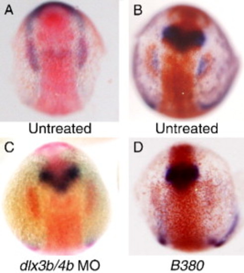
Relationship between dlx3b and sox3 expression. (A) Double in situ hybridization illustrating the appearance of placodal sox3 expression (red) at the caudal end of the pre-placodal domain of dlx3b expression (blue) at the 3-somite stage of development (11 hpf). (B–D) Double in situ hybridization for sox3 expression (red) and pax2a expression (blue) at 12 hpf. In untreated embryos expression of pax2a occupies a domain adjacent to, and medial to sox3 expression (B). Injection of embryos with morpholinos directed against both dlx3b and dlx4b results in loss of pax2a expression in the otic region, but has no effect on sox3 expression (C). Embryos homozygous for the B380 mutation show an identical loss of pax2a expression and normal sox3 expression (D). |
| Genes: | |
|---|---|
| Fish: | |
| Knockdown Reagents: | |
| Anatomical Terms: | |
| Stage Range: | 1-4 somites to 5-9 somites |
| Fish: | |
|---|---|
| Knockdown Reagents: | |
| Observed In: | |
| Stage: | 5-9 somites |
Reprinted from Developmental Biology, 303(2), Sun, S.K., Dee, C.T., Tripathi, V.B., Rengifo, A., Hirst, C.S., and Scotting, P.J., Epibranchial and otic placodes are induced by a common Fgf signal, but their subsequent development is independent, 675-686, Copyright (2007) with permission from Elsevier. Full text @ Dev. Biol.