
Fig. 2
- ID
- ZDB-FIG-111025-44
- Publication
- Sun et al., 2007 - Epibranchial and otic placodes are induced by a common Fgf signal, but their subsequent development is independent
- Other Figures
- All Figure Page
- Back to All Figure Page
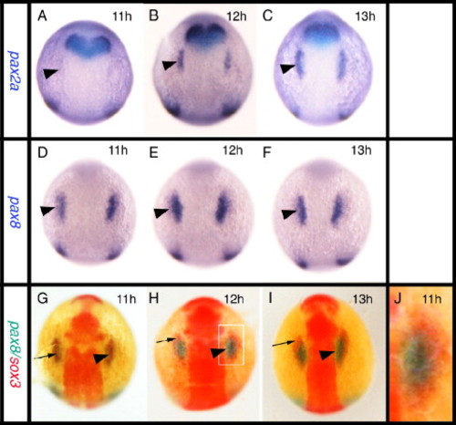
Relationship between the expression of sox3 and the otic markers pax2a and pax8. All panels viewed dorsally at the level of the hindbrain with rostral to the top. In situ hybridization for pax2a (A–C) or pax8 (D–F) expression. (G–J) Double in situ hybridization showing pax8 expression (blue) and sox3 expression (red). pax8 is expressed in the prospective otic placodes (arrowheads) first appearing as discrete domains between 9 and 11 hpf (D) when pax2a is barely detectable in the prospective otic region (arrowhead in A). Double in situ hybridization shows that the initial domain of pax8 expression (arrowhead) is largely coincident with the first expression of sox3 (arrow) in this region. By 12 hpf, however, when pax2a is strongly expressed (B, C), the two domains are almost mutually exclusive with pax8 (and pax2a, see also Fig. 5A) occupying a more medial domain and sox3 an arc of expression surrounding the pax8 expressing ectoderm (G–I). (I) Enlarged view of boxed domain in panel G. |
| Genes: | |
|---|---|
| Fish: | |
| Anatomical Terms: | |
| Stage Range: | 1-4 somites to 5-9 somites |
Reprinted from Developmental Biology, 303(2), Sun, S.K., Dee, C.T., Tripathi, V.B., Rengifo, A., Hirst, C.S., and Scotting, P.J., Epibranchial and otic placodes are induced by a common Fgf signal, but their subsequent development is independent, 675-686, Copyright (2007) with permission from Elsevier. Full text @ Dev. Biol.