|
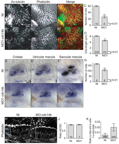
Cdc14B loss-of-function effect on inner ear kinocilia. (A-B′) Saccular macula at 4 dpf visualized with phalloidin and an anti-Ac-tubulin antibody. The latter structure is outlined in blue; the lateral crista is outlined in red. (C,D) Cilia numbers (C) and length (D) were determined at 4 dpf. (E-F′) myo7a in situ hybridization in the inner ear at 4 dpf. (G) The number of cells in the saccular macula was determined in non-injected and MO1-cdc14b-injected larvae at 4 dpf. (H,I) Structure of the utricular macula in non-injected (H) and MO1-cdc14b-injected (I) larvae at 4 dpf visualized by phalloidin. (J) Ratio between the numbers of hair cells (HC) and supporting cells (SC) at 4 dpf. (K) Ratio between the numbers of stereocilia (s.cilia) not associated to kinocilia (k.cilia) and the total number of stereocilia. The numbers of larvae (C,G,J), cilia (D) and hair cells (K) analyzed are indicated. Data are mean ± s.e.m. Statistics were carried out using Student′s t-test.
|