|
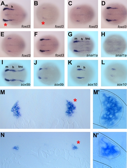
mych MO impairs early neural crest induction.
A–F. Dorsal view of foxd3 expression at 4-somite stage embryos. Embryos injected with 4ng of mych UTR MO (B), 5ng of mych SP MO (C), or 2ng of mych UTR MO (B) plus 2.5ng of mych SP MO (C) showed reduced foxd3 expression. This phenotype was rescued by mych mRNA coinjection (D,F). G–L. Dorsal view of control MO-injected (G,I,K) and mych UTR MO injected embryos (H,J,L) hybridized to snail1a (G,H), sox9b (I,J), and sox10 (K,L). M–N. Sections of the foxd3 expression region of cont MO (M,M′) and mych UTR MO (N,N′) injected embryos. Red asterisks in A and B indicate the location of transverse sections, and asterisks in M and N indicate the regions magnified in M′, N′. h; hindbrain neural crest, m; midbrain neural crest, tnc; trunk neural crest.
|