
- Title
-
Premature Vertebral Mineralization in hmx1-Mutant Zebrafish
- Authors
- El Fersioui, Y., Pinton, G., Allaman-Pillet, N., Schorderet, D.F.
- Source
- Full text @ Cells
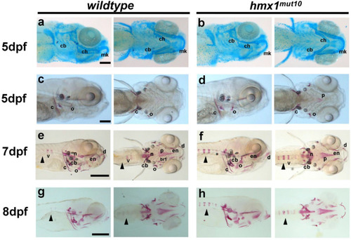
Hmx1mut10 zebrafish embryos develop early mineralized vertebrae: Cartilage structures stained with alcian blue in wildtype and hmx1mut10 embryos (a,b) at 5 dpf; no morphological differences in the developing cranial cartilage structures were detected. Cranial bone structures stained with alizarin red in wildtype and hmx1mut150 at 5 dpf (c,d); wildtype and hmx1mut10 present regular development of bone structures. At 7 dpf and 8 dpf, alizarin red staining detected an early and progressive mineralization of the vertebrae in hmx1mut10 (f,h) in comparison to wildtype zebrafish (e,g). cb, ceratobranchial pairs; ch, ceratohyal; mk, Meckel’s cartilage; v, vertebrae; c, cleithrum; n, notochord; cb, ceratobranchial 5; en, entopterygoid; o, opercle; d, dentary. Black arrowhead: early mineralization of the vertebrae in hmx1mut10 (f,h). Bar, (a–c) 250 μm; (e–g) 500 μm. |
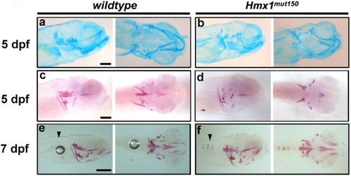
Precocious vertebral bone development in hmx1mut150 zebrafish embryos: Cartilage structures stained with alcian blue in wildtype and hmx1mut150 embryos (a,b) at 5 dpf; the developing cranial cartilage structures develop properly in hmx1-mutant embryos. Cranial bone structures stained with alizarin red in wildtype and hmx1mut150 at 5 and 7 dpf (c,f). Wildtype and hmx1mut150 cranial bones developed regularly at 5 dpf (c,d), while at 7 dpf early mineralization of the vertebrae was detected in hmx1mut150, similarly to hmx1mut10 embryos (e,f). Bar, (a–c) 250 μm; (e–f) 500 μm. |
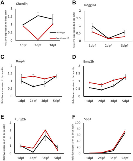
Hmx1mut10 embryos present enhanced expression of osteogenic factors: RT-PCR analysis of the BMP antagonist chordin and noggin1 relative to β-actin, the osteogenic inducers bmp2b and bmp4, and the osteogenic markers runx2b and spp1. In hmx1mut10 embryos, chordin expression is reduced at 2 dpf (A), while noggin1 expression is lower at 1 dpf and 3 dpf (B). Bmp2b and bmp4 transcripts are significantly different in hmx1mut10 embryos when compared to wildtype zebrafish from 1 dpf to 3 dpf (C,D). The osteogenic markers runx2b and spp1 are differentially expressed, and are higher in hmx1mut10 at 2 dpf and 3 dpf, respectively (E,F). Data are expressed as mean of three or more experiments. PHENOTYPE:
|

In situ hybridization detected bmp2b and bmp4 the dorsal region in Hmx1mut10 embryos: In situ hybridization in wildtype and hmx1mut10 embryos at 2 dpf. Staining for bmp2b, bmp4, and runx2b. No signal was detected for bmp2b and bmp4 in the dorsal region of wildtype zebrafish, while bmp4 was detected in the heart region ((A,C) red arrows). In hmx1mut10, bmp2b and bmp4 are expressed in the dorsal region (B,D). Runx2b in wildtype embryos at 2 dpf is expressed in the bone-forming regions (E). In hmx1mut10 embryos, runx2b is expressed in the dorsal region, and presents a similar pattern to bmp2b and bmp4 (F). Cl, cleithrum; ch, ceratohyal; cb, ceratobranchial; h, heart; op, opercle. Bar, A–F 100 μm. |

DMH1-mediated inhibition of BMP signaling reduces the progression of vertebral mineralization: Wildtype and hmx1mut10 embryos were treated with 50 μM DMH1 at 2 dpf and placed in E3 medium at 3 dpf. Alizarin red staining to detect mineralized structures was performed at 7 and 8 dpf (left, lateral view, right ventral view). Wildtype and hmx1mut10 control embryos treated with DMSO (A). Precocious vertebral mineralization was inhibited in DMH1-treated hmx1mut10 embryos at 7 dpf (B). At 8 dpf, treated hmx1mut10 embryos still presented a vertebral mineralization pattern similar to wildtype embryos (B). Bar, (A,B) 500 μm. PHENOTYPE:
|