|
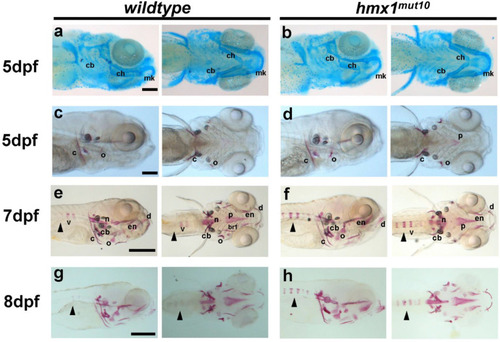
Hmx1mut10 zebrafish embryos develop early mineralized vertebrae: Cartilage structures stained with alcian blue in wildtype and hmx1mut10 embryos (a,b) at 5 dpf; no morphological differences in the developing cranial cartilage structures were detected. Cranial bone structures stained with alizarin red in wildtype and hmx1mut150 at 5 dpf (c,d); wildtype and hmx1mut10 present regular development of bone structures. At 7 dpf and 8 dpf, alizarin red staining detected an early and progressive mineralization of the vertebrae in hmx1mut10 (f,h) in comparison to wildtype zebrafish (e,g). cb, ceratobranchial pairs; ch, ceratohyal; mk, Meckel’s cartilage; v, vertebrae; c, cleithrum; n, notochord; cb, ceratobranchial 5; en, entopterygoid; o, opercle; d, dentary. Black arrowhead: early mineralization of the vertebrae in hmx1mut10 (f,h). Bar, (a–c) 250 μm; (e–g) 500 μm.
|